
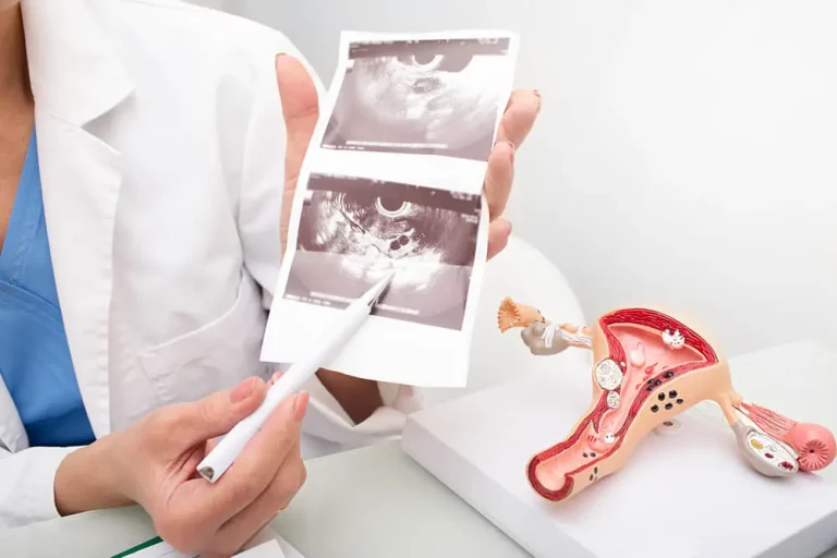
Transvaginal Ultrasound: Everything You Need to Know and Except
A transvaginal ultrasound is a widely performed examination used to identify issues within the female reproductive system. If your doctor has scheduled a pelvic ultrasound, here’s what you can expect.
Why Do Doctors Request a Pelvic Ultrasound?
Doctors may order a pelvic ultrasound for various reasons, including:
- Pelvic pain
- Abnormal uterine or vaginal bleeding
- Checking pregnancy status
- Evaluating the cervix during pregnancy
- Family history of ovarian cancer or other risks like BRCA mutations
We’ll explore these symptoms in detail later.
The Pelvic Ultrasound Procedure: Transabdominal and Transvaginal
An ultrasound, like any medical imaging test, requires a doctor’s prescription if done within the NHS. You can’t simply request one; a medical indication is necessary. However, private clinics like London Private Ultrasound may perform the scan without a referral if you have symptoms.
Preparing for the Ultrasound
Before your ultrasound appointment, you might be instructed to drink water and avoid emptying your bladder. A full bladder helps visualize the uterus and ovaries during a transabdominal scan. In some clinics, you might be offered a transvaginal scan instead.
During the Examination
In the waiting room, the sonographer will call your name. Despite the discomfort from a full bladder, the sonographer will proceed as efficiently as possible. In the exam room, you’ll lie on your back and lower your clothing slightly. The sonographer will apply gel to the transducer and begin scanning your lower abdomen. Inform them if the pressure is too much.
Ultrasound works by sending high-frequency sound waves through the transducer, which then listens for echoes from your tissues. The scan images the uterus and ovaries, noting any abnormalities.
Transvaginal Ultrasound
A pelvic ultrasound typically includes an abdominal scan and a transvaginal ultrasound. The transvaginal probe is longer and thinner than the abdominal one, providing higher resolution images as it gets closer to the uterus and ovaries. The abdominal scan provides an overall view, while the vaginal scan offers a detailed look.
If you haven’t been sexually active, a vaginal ultrasound might not be performed. The vaginal probe will be covered with a condom and disinfected between uses.
Before the Transvaginal Scan
For a vaginal ultrasound, your bladder should be empty. You will be asked to empty your bladder before the scan. If you are using a tampon, you will need to remove it. The sonographer might provide an absorbent pad if you are bleeding heavily. Don’t feel embarrassed; the sonographer’s goal is to help diagnose any issues.
Some exam tables have stirrups for your feet. If not, you might use a pillow or booster pad under your hips or place your feet on a chair.
The sonographer will insert the probe or guide you in doing so. It is not painful and is about the size of a tampon. The sonographer will move the probe to capture images of the uterus and ovaries, using Doppler ultrasound to check blood flow.
After the Scan
Once the necessary images are taken, the sonographer will remove the probe, and you can make yourself comfortable again.
Who is the Sonographer?
In the UK, a sonographer performs the ultrasound. In the USA, they are called technologists. These professionals are highly trained, often holding postgraduate degrees. Previously, sonographers were trained on the job, but now they typically complete accredited university programs.
Common Reasons for Pelvic Ultrasound Scans in Detail
Pelvic Pain
Many women experience pelvic pain at some point. The severity and impact vary. If the pain disrupts your daily life or worsens over time, consult your healthcare provider.
Pelvic pain can have multiple causes, making diagnosis complex. It might be necessary to consider other body systems when identifying the cause. Pain can arise from digestive, urinary, or reproductive issues.
To aid diagnosis, be prepared to answer questions about the onset, duration, severity, and nature of the pain.
Acute Pelvic Pain
Acute pelvic pain occurs over a short period and can indicate a serious problem. It may result from infections or inflammation not necessarily linked to reproductive organs. Other sources include bladder, bowel, or appendix issues, diverticulitis, irritable bowel syndrome, or kidney stones.
Chronic Pelvic Pain
Chronic pelvic pain may be constant or intermittent. Conditions like dysmenorrhea, endometriosis, and adenomyosis often cause it. Surgery or severe infections can lead to adhesions or scar tissue, causing persistent pain.
Ovarian Cysts
Ovarian cysts can cause sharp pain if they leak or bleed. Most small cysts resolve without treatment, but larger cysts may need surgical removal.
Ectopic Pregnancy
An ectopic pregnancy occurs outside the uterus, often in a fallopian tube. It causes pain on one side of the abdomen and may include bleeding. This condition is life-threatening and requires immediate medical attention.
Fibroids
Fibroids are non-cancerous growths in the uterus that can cause pain, pressure, and menstrual abnormalities.
Diagnostic Tests for Pelvic Pain
Diagnosing pelvic pain often involves multiple tests, including ultrasound, CT scans, MRI, and bowel tests. Laparoscopy may be necessary for conditions like endometriosis.
Treatment for Pelvic Pain
Treatment varies based on diagnosis. It can range from medication for infections to hormonal therapies or surgery. Other treatments include heat therapy, muscle relaxants, nerve blocks, and relaxation exercises.
Abnormal Uterine Bleeding
Dysfunctional uterine bleeding (DUB) is a common cause of abnormal bleeding during reproductive years. It occurs when other causes of bleeding have been ruled out.
A normal menstrual cycle lasts 21-35 days, with menstruation for 2-7 days. DUB often results from anovulation, where the corpus luteum fails to form, leading to continuous estrogen production and overgrowth of the endometrium.
Diagnosing Abnormal Uterine Bleeding
Diagnosis involves ruling out other causes. Patients may present with various types of bleeding, including prolonged, irregular, or heavy bleeding.
Transvaginal Ultrasound in Pregnancy
Transvaginal ultrasound is common in early pregnancy. It provides clear images of the uterus, placenta, and fetus. This type of ultrasound is safe for both mother and fetus.
Ectopic Pregnancy Detection
Ectopic pregnancies can be detected with transvaginal ultrasound. Early detection is crucial to prevent life-threatening complications.
Ovarian Cancer Screening
Regular pelvic exams can help detect ovarian cancer early. Transvaginal ultrasound and CA-125 blood tests are used for screening, though they are not always reliable.
Genetic Testing for BRCA Mutations
BRCA mutations increase the risk of breast and ovarian cancers. Genetic counseling and testing can help identify individuals at risk.
Pelvic Pain Treatment
The treatment for pelvic pain varies based on the underlying diagnosis. Here are some common approaches:
- Medications: For urinary tract infections (UTIs) or vaginal infections, antibiotics or other medications may be prescribed. Serious infections like pelvic inflammatory disease (PID) might require pharmacologic treatment in a hospital setting. If a sexually transmitted disease (STD) is diagnosed, treating your partner is essential to prevent reinfection.
- Pain Relief: Menstrual cramps can often be alleviated with anti-inflammatory drugs like ibuprofen, which block the production of prostaglandins responsible for uterine contractions.
- Hormonal Therapies: Hormonal treatments, including oral contraceptives and other hormone types, might be necessary depending on the diagnosis.
- Antidepressants: These can be beneficial for some women as they help break the cycle of pain and depression that often accompanies chronic pelvic pain.
- Surgery: In some cases, surgery might be required. Laparoscopy can be performed on an outpatient basis, while more extensive surgeries, such as a hysterectomy, might necessitate a hospital stay. Your healthcare provider will discuss the risks, benefits, and likelihood of success for each procedure. Hysterectomy is not always the best option, especially for chronic pelvic pain.
- Other Treatments: These include heat therapy, muscle relaxants, nerve blocks, and relaxation exercises. Specific treatments will be used for digestive or urinary conditions if diagnosed.
Determining the exact cause of pelvic pain can be challenging and frustrating, but perseverance is crucial. Even if a specific cause isn’t identified, various treatments can help. Maintaining a good relationship with your healthcare provider is essential to find the most effective treatment.
Abnormal Uterine Bleeding
Dysfunctional uterine bleeding (DUB) is the most frequent cause of abnormal vaginal bleeding in women of reproductive age. It is diagnosed after ruling out other organic and structural causes.
A normal menstrual cycle ranges from 21-35 days, with menstruation lasting 2-7 days and an average blood loss of 35-150 mL (about 8 or fewer soaked pads per day, with no more than 2 heavy days).
Pathophysiology
The menstrual cycle begins with the first day of menses, lasting about 4 days, involving the breakdown and shedding of the endometrium’s functionalis layer. The proliferative (follicular) phase follows, extending from day 5 to day 14, characterized by endometrial proliferation due to estrogen stimulation produced by ovarian follicles under follicle-stimulating hormone (FSH) influence. This phase ends with the peak of estrogen production, triggering the FSH and luteinizing hormone (LH) surge.
Ovulation follows the rupture of the ovarian follicle, releasing the ovum. The secretory (luteal) phase is marked by progesterone and less potent estrogen production by the corpus luteum, lasting from day 15 to day 28. The endometrium’s functionalis layer thickens, and the stroma becomes edematous. Without pregnancy, estrogen and progesterone feedback to the hypothalamus, reducing FSH and LH production. The spiral arteries coil and reduce blood flow, eventually causing the functionalis layer to break down, leading to menses.
Types of DUB
About 90% of DUB cases result from anovulation, while 10% occur with ovulatory cycles. Anovulatory cycles fail to form the corpus luteum, leading to continuous unopposed estradiol production, overstimulating the endometrium, and eventually causing necrosis and excessive uterine blood flow.
In ovulatory DUB, prolonged progesterone secretion causes irregular endometrial shedding, often linked to low estrogen levels around the bleeding threshold. This leads to portions of the endometrium degenerating and spotting.
DUB can be categorized as:
- Menorrhagia: Prolonged (>7 days) or excessive (>80 mL daily) uterine bleeding at regular intervals.
- Metrorrhagia: Uterine bleeding at irregular, more frequent intervals.
- Menometrorrhagia: Prolonged or excessive uterine bleeding at irregular, frequent intervals.
- Intermenstrual Bleeding: Variable amounts of uterine bleeding between regular menstrual periods.
- Polymenorrhea: Uterine bleeding at regular intervals of less than 21 days.
- Oligomenorrhea: Uterine bleeding at intervals of 35 days to 6 months.
- Amenorrhea: No uterine bleeding for 6 months or longer.
Factors and Risks
- Obesity: Often results in irregular menstrual cycles due to non-ovarian estrogen production, leading to prolonged amenorrhea alternating with metrorrhagia or menometrorrhagia.
- Athletes: A high incidence of DUB is seen among female athletes, often related to luteal phase deficiency and inadequate progesterone production.
- Endometrial Cancer: Important to distinguish from DUB; related to estrogen stimulation and endometrial hyperplasia, presenting with postmenopausal bleeding.
Diagnosis and Examination
- Clinical History: Patients may report amenorrhea, oligomenorrhea, menorrhagia, or metrorrhagia. Questions about menstrual regularity, the last menstrual period, reproductive history, and contraceptive use are crucial.
- Medical History: Include inquiries about hypovolemia, diabetes mellitus, hypertension, thyroid disorders, liver disease, medication usage, and alternative medicine modalities.
- Physical Examination: Assess the patient’s volume status and degree of anemia. Perform a pelvic speculum and bimanual examination to identify vaginal bleeding’s etiology, looking for trauma, foreign bodies, structural abnormalities, and signs of hematologic pathology or liver disease.
Causes
Various organic pathologies can present as abnormal vaginal bleeding, including:
- Endocrine Disorders: Hypothyroidism, hyperthyroidism, Cushing disease, liver disease, hypertension, diabetes mellitus, and adrenal disorders.
- Pregnancy-Related Issues: Abnormal bleeding in pregnancy may be reported as unusual in timing, amount, or duration.
- Trauma: Cervical, vulvar, or vaginal trauma.
- Cancer: Carcinomas of the vagina, cervix, uterus, and ovaries.
- Structural Disorders: Functional ovarian cysts, cervicitis, endometritis, salpingitis, leiomyomas.
- Other Factors: Polycystic ovary disease, vaginal infection, polyps, ectopic pregnancy, hydatidiform mole, blood dyscrasias, excessive weight gain, increased exercise, or stress.
Medication-Related Causes:
- Oral Contraceptives: Breakthrough bleeding can occur with inadequate estrogen and progestin doses, missed pills, or drug interactions (e.g., with phenobarbital, carbamazepine, penicillin, tetracycline, trimethoprim-sulfamethoxazole).
- Progestin-Only Compounds: Medroxyprogesterone acetate (Depo-Provera) can cause prolonged uterine breakthrough bleeding. The Norplant system has similar adverse effects.
- Intrauterine Devices (IUDs): Can cause variable vaginal bleeding initially and intermittent spotting subsequently. The progesterone-impregnated IUD (Mirena) is associated with less menometrorrhagia and may lead to secondary amenorrhea.
Transvaginal Ultrasound in Pregnancy
Transvaginal ultrasound has been in use since 1958 and is almost routine in prenatal care. It creates images by “bouncing” sound waves off tissues, using real-time technology to display images on a screen, which can also be recorded digitally, on videotape, or as still photos.
Ultrasound Safety
Ultrasound appears to be completely safe for the fetus, with no demonstrated risks in its 25 years of common use.
Types of Ultrasound
- Trans-Abdominal Ultrasound (TA-US): Most useful in the second and third trimesters. The technician applies gel to your abdomen and moves the transducer over your uterus to create images of the fetus. You may view moving images of your baby on the screen.
- Transvaginal Ultrasound (TV-US): Commonly used in the first trimester and as an adjunct to TA-US later in pregnancy. The TV-US transducer, shaped like a long narrow probe and covered with a condom for sterility, is placed in your vagina. It feels similar to a Pap test. Neither type should be painful, though TA-US might require a full bladder, which can be uncomfortable.
Having an Ultrasound Scan
Obstetrical ultrasound can be performed in two primary ways:
Trans-abdominal ultrasound (TA-US): Most useful in the second and third trimesters. A gel is applied to your abdomen to transmit sound waves, and the technician moves the transducer (which looks like a microphone) over your uterus to create images of the fetus. You can see moving pictures of your baby on the screen.
Transvaginal ultrasound (TV-US): Commonly used in the first trimester and sometimes as an adjunct to TA-US later in pregnancy. The TV-US transducer is shaped like a long, narrow probe, covered with a condom for sterility, and placed in your vagina. Gel is placed in the condom and on its outer surface for sound wave transmission and lubrication. It feels similar to having a Pap test. Neither type should be painful, although a full bladder might be uncomfortable during TA-US.
Who Needs an Ultrasound?
There is ongoing debate about whether ultrasounds should be routine or used only for specific concerns or problems in pregnancy. While research hasn’t shown that routine ultrasounds improve outcomes, many practitioners and parents prefer them for reassurance. Routine ultrasounds are generally considered safe, but financial costs may limit their use.
Reasons for Having an Ultrasound in Pregnancy
- Unclear due date
- Vaginal bleeding or severe abdominal pain
- Abnormal uterus size during prenatal visits
- High risk of poor fetal growth (e.g., twins, maternal obesity)
- Increased likelihood of birth defects
- Any situation where imaging the baby, placenta, uterus, or cervix aids in maternal and fetal care
Transvaginal Ultrasound in Ectopic Pregnancy
An ectopic pregnancy occurs when a fertilized egg implants outside the uterus, often in the fallopian tubes (tubal pregnancy). It can also implant in the ovary, abdomen, or cervix. As the fetus grows, it can cause the organ to burst, leading to severe bleeding and endangering the mother’s life. A classical ectopic pregnancy cannot result in a live birth.
Signs and Symptoms of Ectopic Pregnancy:
- Pain or vaginal bleeding, often sharp and stabbing, possibly in the pelvis, abdomen, shoulder, or neck.
- Vaginal spotting
- Dizziness or fainting due to blood loss
- Low blood pressure
- Lower back pain
Causes of Ectopic Pregnancy:
- Blocked fallopian tubes due to infection or inflammation (e.g., PID caused by gonorrhea or chlamydia)
- Endometriosis or scar tissue from previous surgeries
- Birth defects or abnormal growths altering tube shape
Diagnosis:
- Urine Pregnancy Test: Quick and crucial in emergency cases.
- Quantitative hCG Test: Measures hormone levels, lower than expected levels may indicate ectopic pregnancy.
- Ultrasound Examination: Determines if a fetus is developing in the uterus or if masses are present elsewhere. Pelvic exams can locate pain areas, check for an enlarged uterus, or find masses. Early detection (<5 weeks) is challenging; repeated hCG tests and monitoring may be necessary.
Treatment Options:
- Methotrexate Injection: For early ectopic pregnancy to stop embryo growth.
- Surgery: Required for advanced pregnancies. Laparoscopy is less invasive, involving small incisions and a video camera to remove the ectopic pregnancy. Regular follow-up ensures hCG levels return to zero, indicating no remaining ectopic tissue.
Future Pregnancies:
- Women with a history of ectopic pregnancies may face difficulties conceiving again, especially if they had fertility issues beforehand. Each subsequent ectopic pregnancy increases the risk of recurrence.
Risk Factors:
- Women over 35
- History of PID, previous ectopic pregnancies, fallopian tube surgeries, infertility treatments
- Certain birth control methods (e.g., progesterone-only pills, IUDs, morning-after pill)
- Smoking and multiple sexual partners
When to Call Your Doctor:
- Discuss options if you believe you’re at risk for an ectopic pregnancy before becoming pregnant.
- Use condoms to protect against STDs that can cause PID.
- If pregnant, any pain, bleeding, or ectopic pregnancy symptoms warrant immediate medical attention.
Transvaginal Ultrasound and Ovarian Cancer
Early Detection: About 20% of ovarian cancers are found at an early stage, significantly improving survival rates. Regular pelvic exams can help detect reproductive system cancers early, though early ovarian tumors are often difficult to feel. The Pap test detects cervical cancer but not ovarian cancer.
Symptoms:
- Abdominal swelling or bloating
- Pelvic pressure or pain
- Difficulty eating or feeling full quickly
- Urinary symptoms (urgent or frequent need to urinate)
Screening Tests:
- Transvaginal Ultrasound: Uses sound waves to examine the uterus, fallopian tubes, and ovaries. It can detect masses but can’t distinguish between cancerous and benign masses.
- CA-125 Blood Test: Measures the CA-125 protein, elevated in many women with ovarian cancer. Useful for guiding treatment but not as a screening test due to false positives from other conditions.
Current Screening Recommendations: No major organizations recommend routine use of transvaginal ultrasound or CA-125 for ovarian cancer screening in average-risk women. Screening may be offered to high-risk women with inherited genetic syndromes, but the effectiveness in reducing ovarian cancer mortality is unclear. Ongoing research aims to improve screening tests.
Genetic Testing for BRCA Mutations: BRCA1 and BRCA2 mutations significantly increase the risk of breast and ovarian cancer. Genetic counseling is recommended for those with a family history suggesting a higher likelihood of mutation. Testing can be done via blood or saliva samples, with insurance coverage typically for high-risk individuals. A positive result indicates a predisposition but not a certainty of developing cancer. Negative results in families with known mutations show no BRCA-related predisposition but don’t rule out other genetic factors.
Conclusion
Understanding ultrasound procedures, potential diagnoses, and treatments for pelvic pain, abnormal uterine bleeding, and ovarian cancer is crucial. This knowledge ensures informed discussions with healthcare providers and helps manage expectations for diagnosis and treatment outcomes.
To book online select the date and time that suits you best – alternatively, please contact us with any questions via the chat, call or email links provided.
Telephone: 020 7101 3377