
Doppler Ultrasound Scan: Comprehensive Guide and Applications
A Doppler ultrasound scan is a non-invasive imaging technique that uses high-frequency sound waves to evaluate blood flow through blood vessels. This type of ultrasound provides crucial information about the circulation in your arteries and veins, helping to diagnose various conditions and monitor treatments. Here’s a comprehensive guide on what a Doppler ultrasound scan involves, its uses, preparation, procedure, and what you can expect.
What is a Doppler Ultrasound?
A Doppler ultrasound, also known simply as a Doppler scan, uses the Doppler effect to assess the movement of blood through vessels. Unlike standard ultrasound, which produces images of structures, Doppler ultrasound specifically measures the direction and speed of blood flow.
How Does it Work?
The Doppler effect refers to the change in frequency or wavelength of sound waves in relation to an observer moving relative to the wave source. In medical Doppler ultrasound, sound waves are transmitted into the body, where they bounce off moving blood cells. The returning sound waves are then analyzed to determine the speed and direction of blood flow.
Uses of Doppler Ultrasound
Diagnostic Purposes
Doppler ultrasound is a versatile tool used for diagnosing various conditions related to blood flow and vascular health. Some of the primary diagnostic purposes include:
- Evaluating Blood Flow: Assessing blood flow in arteries and veins to detect blockages, narrowing (stenosis), or blood clots. This is crucial for diagnosing conditions such as deep vein thrombosis (DVT) and peripheral arterial disease (PAD).
- Detecting Peripheral Arterial Disease (PAD): Identifying reduced blood flow in the arteries of the legs or arms. PAD is a common circulatory problem in which narrowed arteries reduce blood flow to the limbs.
- Diagnosing Deep Vein Thrombosis (DVT): Detecting blood clots in the deep veins of the legs. Early detection of DVT is crucial to prevent complications like pulmonary embolism, where a clot travels to the lungs.
- Assessing Varicose Veins: Evaluating the function of valves in veins and detecting varicose veins. This helps in planning treatments such as sclerotherapy, laser therapy, or surgery.
- Monitoring Blood Flow to Organs: Checking blood flow to organs like the kidneys and liver to ensure they are receiving adequate blood supply and functioning properly.
- Pregnancy: Monitoring blood flow in the umbilical cord and placenta to ensure the fetus is receiving adequate blood supply. This is crucial for the health and development of the fetus.
Specialized Uses
Doppler ultrasound has specialized applications that provide detailed insights into specific areas of the body:
-
- Purpose: To assess blood flow in the carotid arteries in the neck, which supply blood to the brain.
- Procedure: The transducer is placed on the neck to visualize the carotid arteries.
- Importance: Detecting narrowing or blockages in the carotid arteries can prevent strokes by allowing for timely intervention.
-
Ankle-Brachial Pressure Index (ABPI)
- Purpose: To compare the blood pressure in the ankle with the blood pressure in the arm.
- Procedure: Doppler ultrasound is used to measure blood flow in both locations.
- Importance: ABPI helps diagnose PAD and assess the severity of the disease.
-
- Purpose: To evaluate blood flow in the arteries of the arms or legs.
- Procedure: The transducer is moved along the arteries to detect blockages or narrowing.
- Importance: Identifies issues like PAD, helping to guide treatment decisions.
-
- Purpose: Specifically focuses on blood flow in the peripheral arteries.
- Procedure: The transducer is moved along the peripheral arteries to identify blockages or reduced flow.
- Importance: Early detection of PAD can prevent complications and guide lifestyle changes and treatments.
-
- Purpose: Comprehensive assessments of vascular health.
- Procedure: May include a combination of Doppler ultrasound scans, ABPI, and other tests.
- Importance: Provides a complete picture of vascular health, identifying risks and guiding preventive measures and treatments.
Differences Between Doppler Ultrasound and Standard Ultrasound
Standard Ultrasound
- Purpose: Standard ultrasound, also known as B-mode or grayscale ultrasound, is primarily used to create images of internal organs and structures. It is commonly used in obstetrics to monitor fetal development, in cardiology to view the heart, and in general medicine to visualize organs such as the liver, kidneys, and gallbladder.
- Imaging: Produces static or real-time images of structures within the body.
- Function: Does not provide information about blood flow. It is used to detect structural abnormalities, tumors, cysts, and other physical changes in tissues and organs.
- Procedure: The transducer emits sound waves that bounce off tissues, creating echoes that are converted into images on a monitor.
Doppler Ultrasound
- Purpose: Doppler ultrasound specifically measures the direction and speed of blood flow through blood vessels. It is used to assess circulatory conditions such as blockages, blood clots, and narrowed arteries.
- Imaging and Sound: Provides real-time imaging combined with auditory signals to represent blood flow. The Doppler effect produces pulsing sounds that correspond to blood flow, which are used to evaluate the speed and direction of blood flow.
- Function: Evaluates blood flow dynamics, helping to diagnose vascular conditions, monitor blood flow to organs, and assess fetal health during pregnancy.
- Procedure: The transducer sends out sound waves that bounce off moving blood cells. The returning sound waves are analyzed to determine the speed and direction of blood flow.
Preparing for a Doppler Ultrasound
Preparation Steps
- Clothing: Wear comfortable, loose-fitting clothing. You may need to remove clothing and wear a gown during the scan.
- Fasting: Depending on the area being examined, you might be asked to fast for a few hours before the procedure. For example, fasting is often required for abdominal Doppler ultrasounds to reduce interference from bowel gas.
- Medications: Continue taking your regular medications unless instructed otherwise by your healthcare provider. Inform your provider of any medications you are taking.
During the Doppler Ultrasound
The Procedure
- Positioning: You will lie on an examination table. The technician (sonographer) will position you to get the best images. For some scans, you might need to lie on your back, side, or stomach.
- Gel Application: A water-based gel will be applied to the skin over the area being examined. The gel helps the sound waves travel through the skin.
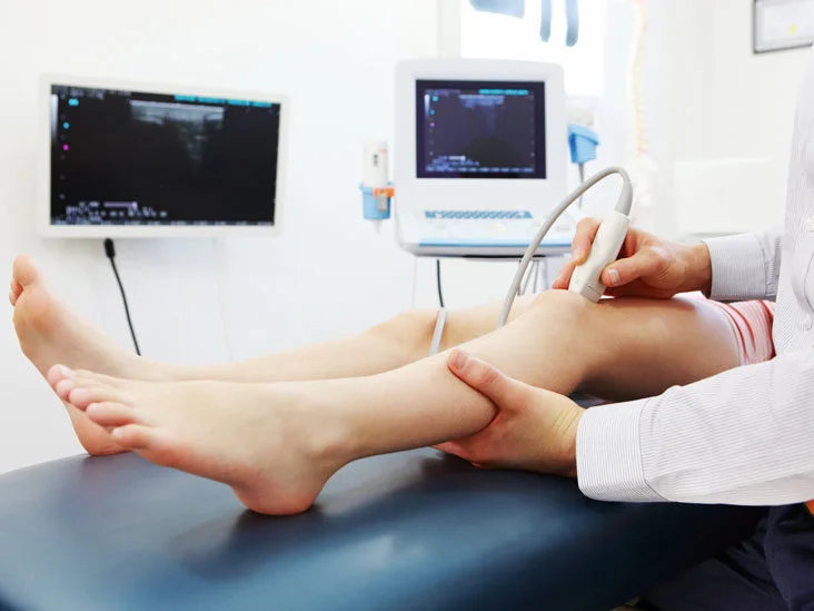
- Transducer: The sonographer will move a handheld device called a transducer over the area. The transducer sends out sound waves and receives the echoes that bounce back.
- Imaging and Sound: As the transducer moves, you may hear pulsing sounds that correspond to the blood flow in your vessels. The sonographer will capture images and measurements during the scan.
- Duration: The scan typically takes 30-60 minutes, depending on the area being examined and the complexity of the test.
What You Will See and Hear
- Monitor: You may be able to see real-time images of your blood vessels on a monitor.
- Pulsing Sounds: The Doppler effect produces sounds that correspond to your blood flow, which the sonographer uses to evaluate the speed and direction of blood flow.
After the Doppler Ultrasound
Discussing the Results
- Immediate Feedback: In many cases, the sonographer can provide initial feedback immediately after the scan.
- Detailed Report: A detailed report will be prepared and sent to your healthcare provider, who will discuss the results with you during a follow-up appointment.
Next Steps
- Further Testing: If any abnormalities are detected, your healthcare provider may recommend additional tests or imaging studies.
- Treatment Plan: Based on the results, your provider will discuss treatment options or necessary interventions to address any identified issues.
Benefits of Doppler Ultrasound
- Non-Invasive and Painless: The procedure is non-invasive, painless, and does not involve exposure to radiation.
- Real-Time Imaging: Provides real-time information about blood flow and vessel condition.
- Versatile: Can be used to evaluate blood flow in various parts of the body, including limbs, neck, abdomen, and during pregnancy.
Risks and Limitations
Risks
Doppler ultrasound is generally safe and has no known risks associated with its use. It does not involve ionizing radiation, making it safe for repeated use and for use during pregnancy.
Limitations
- Image Quality: Image quality may be affected by patient factors such as obesity, gas in the intestines, or movement during the scan.
- Technical Expertise: Accurate interpretation requires a skilled sonographer and radiologist.
Doppler ultrasound is a valuable diagnostic tool for assessing blood flow and detecting vascular conditions. Understanding what to expect during the procedure can help alleviate any anxiety and ensure a smooth experience. The information obtained from a Doppler scan is crucial for diagnosing and managing various health conditions effectively.
To book online select the date and time that suits you best – alternatively, please contact us with any questions via the chat, call or email links provided.
Telephone: 020 7101 3377