
Private ultrasound elastography · Harley Street, Marylebone & Central London · 7 days a week
Private Shear Wave Elastography in London
Shear wave elastography is a quantitative ultrasound elastography technique that measures tissue stiffness in kilopascals (kPa). It is used for the liver, thyroid, breast and testis, and the kPa reading is interpreted alongside your conventional ultrasound images, symptoms, medical history and any previous scans or blood tests. Ultrasound + shear wave elastography starts at £499, breast ultrasound + shear wave elastography is £539, and a standalone SWE add-on is available from £360 if you have a recent suitable ultrasound report. You do not need a GP referral, and same-day appointments are usually available at 27 Welbeck Street, in the Harley Street medical district.
Shear wave elastography adds a stiffness reading to a conventional ultrasound. The kPa result is read together with the greyscale images, your symptoms, your history and any previous scans, so the report tells you what the number means in your clinical context.
- Ultrasound + SWE from £499
- Standalone SWE from £360
- Quantitative kPa readings
- Liver, thyroid, breast & testis
- Written report within 24 hours
- No GP referral required
Tell us the organ (liver, thyroid, breast or testis) and the reason for the scan (a raised liver enzyme, a nodule, a lump, a previous kPa reading you would like repeated), and we will tell you which bundle fits, or whether a standalone shear wave elastography add-on to your existing ultrasound is enough. Call 020 7101 3377 or message us on WhatsApp before you book.
Shear Wave Elastography at a Glance
| What it is | An ultrasound technique (SWE / Shearwave Elastography) that measures tissue stiffness in kilopascals (kPa), performed as part of a conventional ultrasound. |
|---|---|
| Organs covered | Liver, thyroid, breast and testis. Each SWE bundle includes a full conventional ultrasound of the same organ first, then the elastography measurement. |
| Cost | From £499 for ultrasound + SWE (thyroid, liver, testis) · £539 for breast · standalone SWE add-on from £360 where a recent suitable ultrasound report is available. |
| Timing & report | Appointment usually 20–30 minutes. Verbal findings explained in the room. Written report normally sent within 24 hours. For liver SWE a 2-hour fast is recommended (water is fine). |
| Location | 27 Welbeck Street, London W1G 8EN · Harley Street medical district, Central London. SWE is not currently available at the St Albans clinic. |
Organ-based Shear Wave Elastography Packages
Four bundled pathways (liver, thyroid, breast, testis) and a standalone SWE add-on. Every bundle includes a conventional ultrasound of the same organ first, then the elastography measurement where clinically appropriate, then a written report that ties the two parts together. Not sure which one fits? Call 020 7101 3377 before booking.
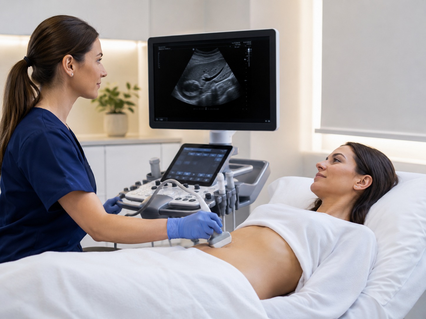 Liver & Fibrosis
Most Booked
Liver & Fibrosis
Most Booked
Liver Ultrasound + Shear Wave Elastography
For liver stiffness assessment and fibrosis screening pathways, including fatty liver, raised liver enzymes on a previous blood test, viral hepatitis follow-up, or serial monitoring of a known finding.
- Conventional liver ultrasound + 2D SWE
- Quantitative kPa stiffness measurement
- Manufacturer-appropriate liver reference values
- 2-hour fast recommended, water permitted
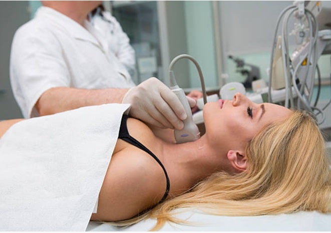 Thyroid Nodules
Thyroid Ultrasound + Shear Wave Elastography
For selected thyroid nodules where kPa stiffness data may support the greyscale and Doppler ultrasound findings, or where SWE has been suggested by a previous imaging report or a referring clinician.
- Full thyroid and cervical node ultrasound
- Point or 2D SWE stiffness measurement
- Interpreted alongside TI-RADS features
- Verbal explanation in the scan room
 Breast Lesions
Breast Ultrasound + Shear Wave Elastography
For selected breast lesions where kPa stiffness data may support the conventional breast ultrasound interpretation. Used alongside BI-RADS assessment, with mammography, MRI and biopsy reserved for cases where those are clinically indicated.
- Bilateral or unilateral breast ultrasound
- Shear wave elastography of the target lesion
- Read alongside BI-RADS features
- Female clinician available on request
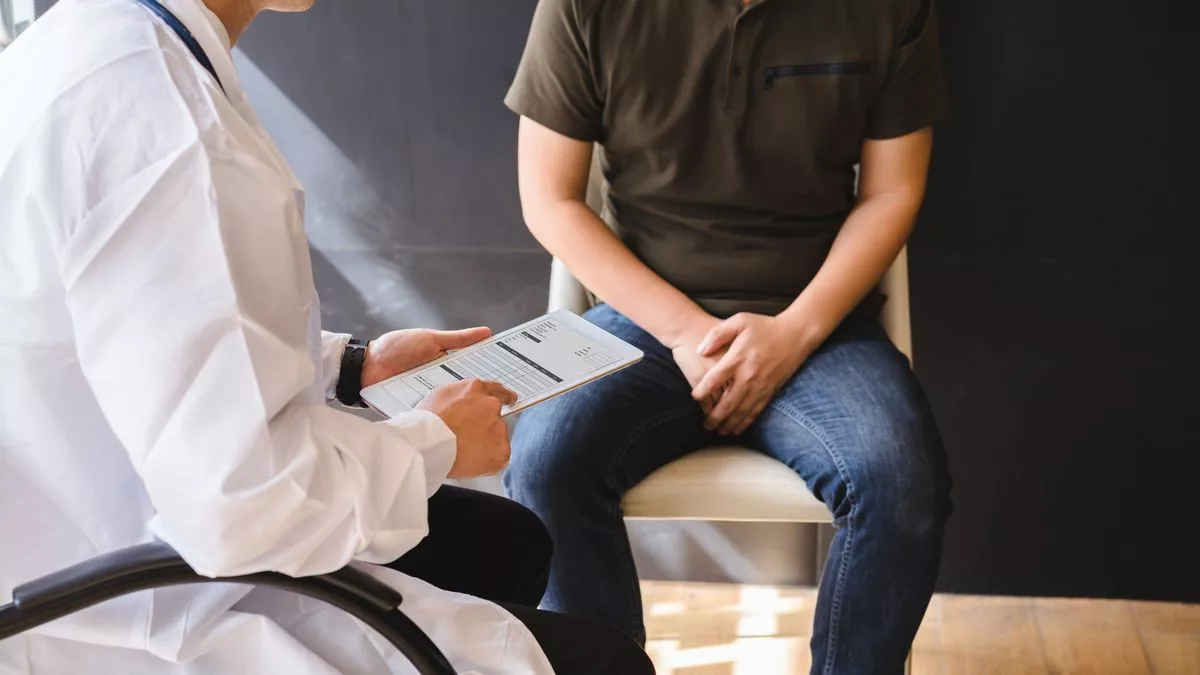 Testis & Scrotum
Testicular Ultrasound + Shear Wave Elastography
For selected testicular or scrotal findings where a kPa stiffness measurement may support the standard testicular ultrasound assessment, or where SWE has been requested by a specialist for follow-up of a known finding.
- Bilateral testicular / scrotal ultrasound
- Shear wave elastography of the target area
- Read alongside greyscale and Doppler findings
- Verbal explanation in the scan room
Add-on
Bring your report
Standalone Shear Wave Elastography
If you already have a recent, clinically suitable conventional ultrasound report for the same organ (from us or elsewhere), a standalone SWE measurement can be added on without repeating the full ultrasound. Subject to clinical suitability at the time of booking.
- Elastography measurement only
- Recent ultrasound report required
- Liver, thyroid or testis: £360
- Breast standalone: £400
Every price includes the appointment, the SWE measurement, a verbal explanation in the room and the written report. The kPa reading is one input into the clinical picture, and the report says exactly what it does and does not tell you. Biopsy, blood tests, mammography, MRI, CT and specialist assessment are used where they are clinically indicated.
How to Prepare for Your Shear Wave Elastography Scan
A shear wave elastography appointment feels no different from a normal ultrasound. Four short things worth doing before you arrive.
Bring your previous imaging and reports
Any previous ultrasound, MRI, CT, biopsy report, kPa reading or specialist letter you have. Serial SWE is only useful if the sonographer can put today’s number next to the last one, and the images give context to the stiffness reading.
Fast for 2 hours if this is a liver scan
For liver shear wave elastography, a 2-hour food fast is recommended and water is fine. The rest of the SWE pathways (thyroid, breast, testis) do not need fasting. Take your usual medication as normal unless a clinician has told you otherwise.
Wear something easy to move aside
You will only need to expose the area being scanned. A two-piece outfit is easier than a dress. A chaperone is available on request, and a female clinician can be requested at booking for breast or pelvic examinations.
Allow about 30 minutes
Most bundled ultrasound + SWE appointments take 20 to 30 minutes. You will get a verbal explanation of the findings in the scan room before you leave, and the written report is usually sent within 24 hours.
Your Shear Wave Elastography Report
Your report is written in plain English. It sets out the conventional ultrasound findings, the SWE kPa measurement where one was obtained, any technical limitations of that number, and a recommendation for what to do next. The pathway from that recommendation follows one of four routes.
Reassuring findings
Where the ultrasound looks unremarkable and the SWE kPa reading sits within the expected range for that organ, the report explains the numbers, gives you self-care or lifestyle advice where relevant, and suggests routine follow-up only if symptoms change. No further imaging is usually needed.
Indeterminate or borderline
Where findings are mixed or the kPa reading is at the edge of the expected range, the report will usually recommend GP or specialist review, targeted blood tests, or a repeat scan in a defined interval to check whether the stiffness is stable, improving or worsening over time.
Suspicious findings
Where features on the ultrasound or the SWE reading raise a specific clinical concern, the report will recommend appropriate onward care through your GP, an NHS specialist pathway (for example a rapid access clinic) or a private specialist. Where relevant we can refer directly.
Urgent care
Where a serious, time-sensitive concern is identified during the appointment (for example, testicular torsion, acute liver decompensation, a suspected acute abdomen), the sonographer will advise you to call 999 or attend A&E immediately rather than wait for the written report.
How we use shear wave elastography safely. Every kPa reading on this pathway is quality-controlled by the reporting clinician against the manufacturer-appropriate reference values for the machine used, and interpreted with the greyscale images, your history and any previous results. Where SWE would add useful information to your ultrasound, it is included in the report; where it would not, we say so plainly and do not charge for it.
The Team Behind Your SWE Scan
Your scan is performed and reported by our clinical ultrasound team, with clinical governance and editorial oversight signed off by a GMC-registered medical director. Say when you book if you would prefer to see a female clinician.
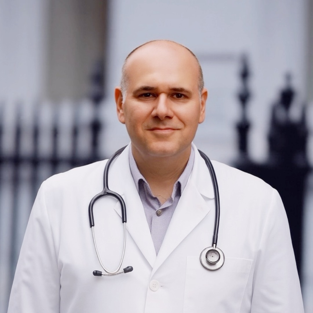
Dr Babak Soleimanpour
Medical Director & GPMD · MRCGP · DRCOG · GMC 6060555 · clinical governance for the shear wave elastography pathway and sign-off on onward referral

Mr Pedram M. Aghaei
Co-founder & Medical WriterWebsite content editing, patient information governance and reporting standards for the elastography clinic

Mrs Fatemeh Selahi
Co-founder & CQC ChampionNurse prescriber · NMC registered · clinic standards, patient safety and CQC compliance
Reporting clinician
Scan & report sign-offYour SWE scan is performed and reported by our clinical ultrasound team. Your written report is signed by the reporting clinician who reviewed the images and stiffness measurements.
Shear Wave Elastography in Harley Street, Marylebone and Central London
Our shear wave elastography service runs from 27 Welbeck Street, W1G 8EN, in the heart of the Harley Street medical district. That puts a private SWE appointment within a short walk of Bond Street, Oxford Circus, Marylebone, Baker Street and Mayfair, and inside a direct journey of Paddington, Victoria, King’s Cross and most of Central London. Same-day and next-day appointments are usually available, 9am to 7pm, seven days a week. Shear wave elastography is not currently offered at the St Albans clinic.
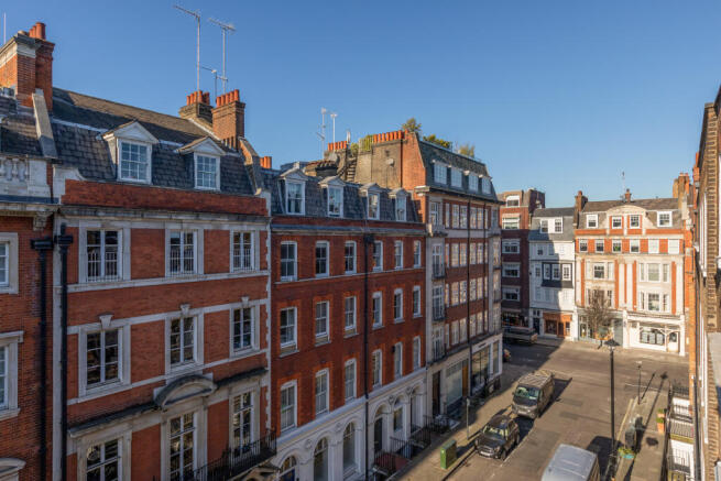
Central London: Harley Street medical district
27 Welbeck Street, London W1G 8EN
Two minutes from Bond Street on the Elizabeth, Central and Jubilee lines, five from Oxford Circus and eight from Marylebone and Baker Street. Convenient for patients travelling in from Marylebone, Fitzrovia, Mayfair, Soho, Chelsea, Knightsbridge, Paddington, South Kensington and wider Central London.
- Harley Street
- Marylebone
- Central London
- Bond Street
- Oxford Circus
- Mayfair
- Fitzrovia
- Soho
- Baker Street
- Paddington
- Chelsea
- Knightsbridge
- South Kensington
- Victoria
- The City
- West End
Shear Wave Elastography London: Frequently Asked Questions
The questions people ask before booking a private SWE scan, including the ones where the honest answer is that we are not the right clinic for the request.
What is shear wave elastography?
Shear wave elastography, often shortened to SWE or Shearwave Elastography, is an ultrasound technique that measures how stiff a tissue is. During a normal ultrasound the machine sends a short focused push into the tissue, then listens for the shear waves that ripple out sideways from that push. The speed of those waves is turned into a stiffness value, reported in kilopascals (kPa).
At London Private Ultrasound we use SWE alongside conventional ultrasound shear wave elastography imaging for the liver, thyroid, breast and testis, and the kPa result is read together with the greyscale images, your symptoms, medical history and any previous scans.
How does shear wave elastography work?
The probe sends a small acoustic radiation force push into the tissue you are being scanned for, which sets off tiny shear waves at right angles to the ultrasound beam. Softer tissue lets those waves travel more slowly, stiffer tissue lets them travel faster, and the machine calculates a stiffness value from the wave speed.
It reports that number in kilopascals (kPa) and, on 2D shear wave elastography, colour-codes the region so the sonographer can see the stiffness pattern across the whole area rather than a single point. You will not feel the push, and nothing about the appointment feels different from a normal ultrasound.
What is the difference between 2D shear wave elastography and point shear wave elastography?
Both are quantitative ultrasound elastography techniques that report tissue stiffness in kPa. Point shear wave elastography (pSWE) measures stiffness from a single small region of interest, one location at a time. 2D shear wave elastography samples stiffness across a larger area at once and displays it as a colour map, which helps the sonographer position the region of interest away from vessels, ribs and other artefacts.
In practice, 2D SWE is what most modern liver, thyroid, breast and testicular protocols now use, because it gives more measurement flexibility and better quality control. Which technique your scan uses depends on the machine and the organ, and both are covered by the SWE bundles on this page.
What is the difference between shear wave elastography and FibroScan?
FibroScan (transient elastography) and shear wave elastography both estimate liver stiffness in kPa, but they use different technology and are not always interchangeable. FibroScan is a dedicated bedside device that uses a mechanical vibration through a single probe and does not show you a live ultrasound image, so measurements are taken blind to the internal anatomy.
Shear wave elastography is built into a full ultrasound machine, so the sonographer sees the liver on the screen, avoids ribs and vessels, and can pick a clean measurement box in real time. SWE also lets us look at the thyroid, breast and testis in the same appointment, which FibroScan cannot. For most patients on our pathway, a liver ultrasound + SWE at £499 is preferred over FibroScan, because you get a full liver ultrasound and the stiffness measurement together, in one visit, from one clinician.
How much does private shear wave elastography cost in London?
At London Private Ultrasound, thyroid, liver and testicular ultrasound bundled with shear wave elastography are £499 each. Breast ultrasound + SWE is £539. Standalone shear wave elastography, without a fresh conventional ultrasound, is available from £360 (breast standalone £400) and is only offered if you already have a recent, clinically suitable ultrasound report for us to compare against.
Every price includes the appointment, the SWE kPa measurement, a verbal explanation in the scan room and a written report usually sent within 24 hours. Compare what is actually in the price rather than the headline number when you are looking at shear wave elastography cost quotes elsewhere, because standalone measurements without a reporting clinician are not the same product.
Do I need a GP referral for shear wave elastography?
No. You can book directly online, by phone on 020 7101 3377, or on WhatsApp. Bring any previous imaging, blood tests, biopsy reports or specialist letters with you if you have them, because they help the sonographer read your SWE result in the right clinical context.
If you have already had a conventional ultrasound elsewhere in the last few months, mention it at booking, because a standalone SWE add-on may be enough rather than a full repeat scan.
Which organs do you offer shear wave elastography for?
London Private Ultrasound offers shear wave elastography for the liver, thyroid, breast and testis, each combined with a conventional ultrasound of the same organ.
Musculoskeletal SWE (for muscle, tendon and other soft tissue stiffness assessment) and prostate shear wave elastography are not currently offered as bookable pathways. If you have been asked to have SWE of an organ outside our current list, call the clinic and we will tell you honestly whether we can help or point you at a service that can.
What ultrasound machine do you use for shear wave elastography?
Our shear wave elastography machine is a premium ultrasound platform from one of the major manufacturers. GE, Canon, Philips and Siemens all make clinically validated SWE systems (GE shear wave elastography, Canon shear wave elastography and Philips shear wave elastography are used interchangeably in the literature), and the platforms are broadly comparable for the organs on this page.
The kPa cut-offs the reporting clinician uses are manufacturer-appropriate, which matters, because thresholds published for one platform should not be applied blindly to results from a different one. If your previous imaging was on a specific machine, tell us at booking so we can note the platform in your report.
What are the normal shear wave elastography values for the liver?
Broadly, most healthy adults produce a liver SWE reading under about 6 kPa, with published shear wave elastography liver normal values varying slightly by manufacturer and technique. Higher values suggest progressively more fibrosis, and the ranges commonly quoted correspond to Metavir stages F0 to F4.
The exact numbers depend on the platform, the sample quality, whether you fasted and whether the underlying cause is fatty liver, viral hepatitis or something else. Because of that, your report explains the number in the context of the images and your history rather than treating one kPa reading as a diagnosis on its own. Any cut-offs mentioned in the report are the manufacturer-appropriate ones for the machine used.
How is shear wave elastography used in the liver and for fatty liver?
Liver shear wave elastography is used to estimate liver stiffness in kPa, which correlates with the degree of fibrosis. In fatty liver disease (including MASLD and previously NAFLD), the ultrasound picks up the fatty change on the greyscale image and SWE adds a stiffness reading that helps flag whether liver fibrosis is likely to have progressed.
In viral hepatitis and other chronic liver conditions, serial SWE is used to monitor whether stiffness is stable, improving or getting worse over time. SWE does not replace liver blood tests, hepatology review or biopsy when those are clinically indicated; it sits alongside them, and the report says exactly what it does and does not tell you.
How is shear wave elastography used for breast lesions?
For selected breast lesions, shear wave elastography of the breast adds a stiffness reading in kPa that can help interpret an ultrasound finding, because malignant lesions tend to be stiffer than benign ones. Radiology practice uses SWE as an adjunct to greyscale and Doppler ultrasound, not as a replacement for BI-RADS assessment, biopsy, mammography or MRI.
On our pathway, breast SWE is only used where the reporting clinician judges it will add useful information to the conventional ultrasound; where it does not, we say so and do not charge you for a measurement that would not change your report. The service does not replace the assessment you would receive at a one-stop breast clinic where biopsy is clinically appropriate.
Do you offer prostate shear wave elastography?
No. London Private Ultrasound does not currently offer prostate shear wave elastography. The clinical evidence for prostate SWE and the transrectal pathway it usually requires sit inside specialist urology practice rather than a general elastography clinic, and we would rather refer that assessment out than overpromise on it here.
If you are looking for a prostate assessment, our private urology clinic and prostate MRI referral pathway are the sensible starting points instead.
Do you offer musculoskeletal or muscle shear wave elastography?
Not as a standalone bookable pathway. Musculoskeletal SWE (for skeletal muscle, tendons and other soft tissues) is an active research area, and the kPa cut-offs and reproducibility for MSK SWE are still being defined in the literature.
For clinical MSK ultrasound (shoulder, knee, hip, hand and wrist, foot and ankle, sport injuries) we use conventional greyscale and Doppler ultrasound through our musculoskeletal clinic. If a specific research or specialist-referred MSK SWE question is being asked, please call the clinic first so we can check what is technically possible before you book.
What are the limitations of shear wave elastography?
SWE reports a stiffness value in kPa, which is one input into the clinical picture. Readings can be affected by patient factors (a fatty liver’s steatosis level, food or alcohol in the last few hours, muscle tension, obesity, ascites, heart failure or inflammation), by measurement technique, by the machine used and by the reference values applied.
Manufacturer-to-manufacturer cut-offs are not always interchangeable, and a single unusually high or low reading in a noisy dataset should not drive a decision on its own. Those are the advantages and disadvantages of shear wave elastography in practice: quantitative and reproducible when done well, but sensitive to context. SWE also does not replace biopsy, mammography, MRI, CT, blood tests or specialist review where those are clinically indicated. Your report explains where SWE added information and, where it did not, says so plainly.
Is shear wave elastography safe, and can I have it if I am pregnant?
Shear wave elastography uses the same diagnostic ultrasound technology as a standard ultrasound scan. There is no ionising radiation, no contrast injection, no biopsy and no needle involved.
If you are pregnant, tell the clinic and the scanning clinician at booking. SWE is not routinely used during pregnancy, and the clinical team will confirm whether the requested scan is appropriate for your situation before it goes ahead. In general, SWE is well tolerated and the appointment feels no different from a normal ultrasound.
How long does the appointment take, and when will I get my report?
Most bundled ultrasound + shear wave elastography appointments take 20 to 30 minutes, depending on the organ and the complexity of the findings. You will get a verbal explanation in the scan room, and a written report is usually sent within 24 hours.
The report includes the conventional ultrasound findings, the kPa measurements where they were obtained, any technical limitations relevant to those numbers, and recommendations for onward care. Same-day and next-day appointments are usually available at 27 Welbeck Street, 9am to 7pm, seven days a week.
This page is for general information and does not replace individual medical advice. Biopsy, blood tests, mammography, MRI, CT, histology and specialist review are used where they are clinically indicated. Seek urgent medical attention rather than booking a routine appointment for sudden severe testicular pain, a painful swollen scrotum with fever, severe abdominal or pelvic pain, jaundice, vomiting blood, heavy vaginal bleeding, collapse, confusion or rapidly worsening symptoms.