

An abdominal ultrasound is one of the most frequently requested scans in UK healthcare, and one of the least explained.
Most people arrive knowing only that they have been told to fast and that gel will be involved.
This guide covers what the scan looks at, organ by organ, what it can and cannot find, how to prepare properly, and how to make sense of the report afterwards.
What is an abdominal ultrasound scan?
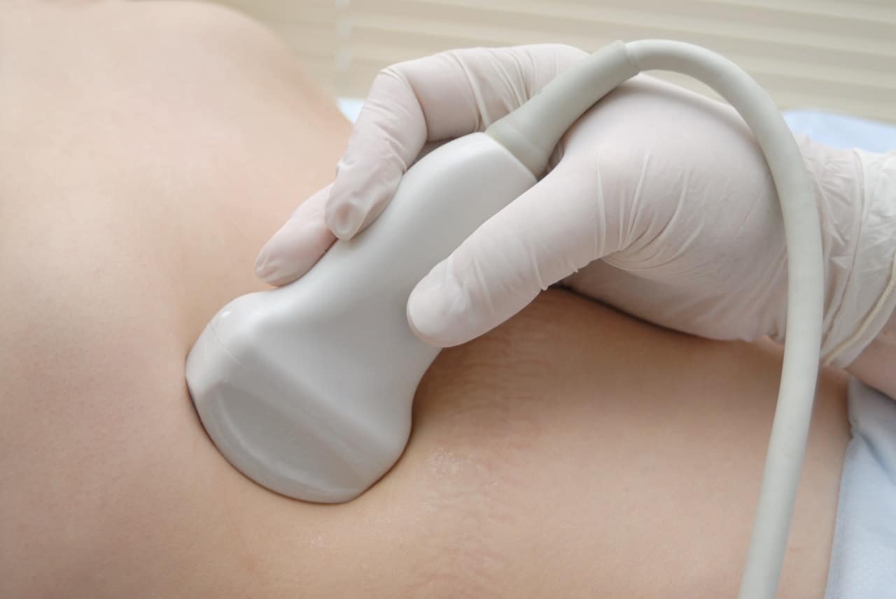
An abdominal ultrasound uses high-frequency sound waves to build real-time images of the organs in your upper abdomen. A handheld probe, called a transducer, is moved across the skin with a layer of gel between the two. The gel removes the air gap that would otherwise stop the sound waves reaching your organs.
The scan involves no ionising radiation and no injections, and nothing enters the body. It is painless, though you may feel firm pressure from the probe over a tender area.
While it is sometimes called a stomach scan or stomach ultrasound, that name can be a bit misleading, as the scan assesses the organs surrounding the stomach rather than the stomach itself.
What does an abdominal ultrasound show?
An upper abdominal ultrasound gives a detailed look at several key organs in your abdomen. Rather than focusing on just one area, it helps build an overall picture of how things are functioning and whether anything looks out of the ordinary.
At LondonSono, we review six main structures as part of a standard scan, and each is reported on individually:
Liver
We assess the size of your liver, its surface, and how it reflects sound waves (called echogenicity). This helps us identify changes such as fatty liver, which often appears brighter on ultrasound—sometimes described as a “bright liver.”
We can also detect common findings like simple cysts, haemangiomas, and features that may suggest chronic liver disease.

Where liver disease is more advanced, ultrasound can show changes that point towards cirrhosis. These include a liver that appears smaller than expected, an irregular or nodular surface, and a coarse internal texture. The scan may also pick up related changes elsewhere, such as an enlarged spleen or fluid in the abdomen, which can develop as pressure builds in the liver’s blood supply.
Cirrhosis happens when long-term inflammation gradually replaces healthy liver tissue with scar tissue. Common causes include fatty liver disease, long-term alcohol use, and hepatitis B or C.
Ultrasound is often the test that first raises the possibility, but it is not used to confirm the diagnosis on its own. That usually involves blood tests, elastography, and in some cases a biopsy.
Gallbladder and bile ducts
This part of the scan looks for gallstones, sludge, or signs of inflammation, and measures the bile ducts to check for any widening.
Ultrasound is the first-line test for suspected gallstones under NICE guidelines. However, results can be affected by preparation—if you’ve eaten beforehand, the gallbladder may be contracted, which can make smaller stones harder to detect.
Also check: Gallbladder Health Package at LondonSono
Pancreas
We examine the head, body, and tail of the pancreas, as well as the pancreatic duct, as far as visibility allows.
In some people, bowel gas can obscure the pancreas, meaning we may not get a complete view. If symptoms persist and the pancreas cannot be clearly assessed, a CT or MRI scan is usually the next step.
Kidneys
Both kidneys are measured and assessed for their structure. We look at the thickness of the outer layer, check for swelling in the drainage system, and note any cysts or masses.
Ultrasound commonly identifies kidney stones, simple cysts, and signs of long-standing kidney conditions. Very small stones can sometimes be missed, which is why a dedicated urinary tract scan may be suggested if needed.
Spleen
We measure the spleen and assess its internal appearance. An enlarged spleen (typically over 12 cm) can be linked to infections, blood disorders, or increased pressure in the liver’s circulation.
Abdominal aorta
The abdominal aorta, the body’s main blood vessel, is measured at several points to check for widening. A diameter of 3 cm or more is considered an abdominal aortic aneurysm.
This is included as a routine safety check rather than a full screening test. In the UK, men are invited for dedicated NHS screening in the year they turn 65.
What an abdominal ultrasound cannot show
Understanding what an abdominal ultrasound cannot show is just as important as knowing what it can. It helps set the right expectations and ensures you are guided toward the most appropriate test if needed.
The bowel
Ultrasound is not a reliable way to assess the bowel in detail. If your main symptoms include a persistent change in bowel habits, rectal bleeding, or unexplained weight loss, this scan is unlikely to provide clear answers.
In these cases, your GP may recommend other tests such as a colonoscopy or CT scan, which are better suited to evaluating the bowel.
Pelvic organs
A standard abdominal ultrasound does not include the pelvic organs. This means structures like the uterus, ovaries, bladder, and prostate are not fully assessed.
If your symptoms relate to these areas, a pelvic or urinary tract scan is usually more appropriate. In many cases, this can be arranged as part of the same visit.
Adrenal glands
The adrenal glands sit deep in the abdomen and are often not visible on ultrasound. Because of their size and position, CT or MRI scans are much more reliable for assessing them.
Diagnosing cancer
Ultrasound is a useful first step and can detect abnormalities such as masses or unusual changes in organs. It can also help indicate whether something appears more or less suspicious.
However, it cannot provide a definitive cancer diagnosis. Confirming the nature of a finding typically requires further imaging and, in many cases, a biopsy.
A normal ultrasound result is reassuring, but like any test, it cannot rule out every possible condition.
Why your doctor might request an abdominal ultrasound
An abdominal ultrasound is commonly requested for:
- Pain in the upper abdomen, particularly under the right ribs
- Pain after fatty meals, or indigestion that has not settled
- Abnormal liver function blood tests
- Abnormal kidney function blood tests
- Jaundice, dark urine or pale stools
- Persistent bloating or a feeling of fullness
- Flank pain, or blood in the urine
- Monitoring a finding identified on a previous scan
How to prepare for an abdominal ultrasound

Preparing properly for your abdominal ultrasound makes a real difference to how clear and accurate your images are. In fact, inadequate preparation is one of the most common reasons a scan needs to be repeated, so it is worth getting this right.
Fast for 4 to 6 hours
Avoid all food and drinks except water during this time. This includes milky drinks and anything sugary. Fasting keeps your gallbladder full, which makes it much easier to detect gallstones, and also helps reduce bowel gas that can interfere with the scan.
Drink plain water
You can continue to have small sips of water throughout the fasting period. Staying hydrated can improve visualisation of the kidneys and generally helps with image quality.
Avoid alcohol for 24 hours
Alcohol can cause the gallbladder to contract, which may make smaller stones harder to see on the scan.
Skip fizzy drinks and chewing gum on the day
Both can introduce extra gas into the digestive system. Bowel gas is one of the main reasons certain organs, especially the pancreas, are not clearly visible on ultrasound.
Take your usual medication
You can take your regular medications with water unless your doctor has advised you otherwise. If you are unsure, it is always best to check in advance.
If you have diabetes, let the clinic know
Let the clinic know when booking your appointment. You may be given an earlier slot to make fasting easier. It is also a good idea to bring something to eat with you for immediately after the scan.
Wear comfortable clothing
Loose, two-piece clothing makes it easier to access the upper abdomen and helps keep the appointment quick and comfortable.
What happens during the abdominal scan
An abdominal ultrasound is a straightforward and non-invasive test, and most people find it quick and comfortable. Here is what to expect:
Before the scan
Your sonographer will begin by asking a few questions about your symptoms, any previous scans, and relevant blood test results.
This context is important because it helps them focus on specific areas and interpret what they see more accurately.
If you have had imaging done before, it is helpful to bring the reports with you. Comparing past and current findings can make a meaningful difference in how results are assessed.
During the scan
You will be asked to lie on your back with your upper abdomen exposed. A warm gel is applied to the skin, which helps the ultrasound probe make good contact and produce clear images.
The sonographer will move the probe gently across different areas of your abdomen, sometimes applying light pressure to get a better view. You may be asked to turn onto your side for certain parts of the scan.
At times, you will be asked to take a deep breath and hold it for a few seconds. This helps bring organs like the liver and gallbladder into clearer view below the rib cage.
How long it takes
The scan itself usually takes around 20 minutes. It is a good idea to allow a bit of extra time for your appointment overall, as there will be some discussion before and after the scan.
After the scan
Once the scan is complete, the gel is wiped off, and you can return to your normal routine straight away, including eating and drinking. There is no downtime or recovery needed.
Understanding your abdominal ultrasound report

Ultrasound reports are written for clinicians, so it is completely normal if the wording feels technical or unclear at first glance.
Understanding a few common terms can make your report much easier to follow and help you interpret what has been found.
Echogenicity
This describes how tissues reflect sound waves on the scan.
- Hyperechoic means brighter than the surrounding tissue. This is how fat, fibrous tissue, and stones often appear.
- Hypoechoic means darker.
- Anechoic means completely black, which usually indicates fluid. This is why simple cysts are typically easy to recognise and are often benign.
Acoustic shadowing
This refers to a dark area seen behind a dense structure that blocks the ultrasound waves. Gallstones and kidney stones commonly produce this effect. In many cases, it is the presence of this shadow that helps confirm the diagnosis rather than the stone itself.
Hepatic steatosis
This means fat in the liver, often referred to as fatty liver. It is a common finding and is usually assessed alongside your clinical history and blood tests.
Hydronephrosis
This describes swelling in the kidney’s drainage system, typically caused by urine not flowing properly. It can be temporary or may need further evaluation depending on the cause.
Splenomegaly
This simply means an enlarged spleen. The significance depends on how enlarged it is and your overall clinical picture.
“Unremarkable” or “within normal limits”
Both of these phrases mean that no abnormalities were detected in that particular organ or structure.
“Limited by bowel gas” or “suboptimal views”
These phrases can sound concerning, but they do not mean that something abnormal was found. They indicate that a particular area could not be seen clearly enough to assess.
When this happens, the report will usually suggest whether further imaging, such as a CT or MRI scan, is needed for a clearer view.
Ultrasound compared with CT and MRI
When your doctor recommends imaging, it is natural to wonder how ultrasound compares with other scans like CT or MRI. Here is a quick overview of what each scan or test tells us:
| Feature | Ultrasound | CT Scan | MRI Scan |
|---|---|---|---|
| Radiation | None | Yes | None |
| Best for | Gallbladder, liver texture, kidneys, aorta | Bowel, pancreas, trauma, cancer staging | Bile ducts, liver lesion characterisation, detailed soft tissue |
| Contrast injection | Not usually required | Often required | Often required |
| Typical scan time | About 20 minutes | Around 10 minutes | 30 to 45 minutes |
| Main limitation | Visibility reduced by bowel gas or body habitus | Involves radiation exposure | Higher cost, limited availability, enclosed scanner |
What a private abdominal ultrasound costs in the UK
Private abdominal ultrasound in London generally runs from about £180 to £250, depending on the clinic, what is included and how quickly the report is delivered. Check three things before comparing prices:
- Whether the consultation with the sonographer is included or billed separately
- Whether the written report is included, and how quickly it arrives
- Which organs are covered, since some clinics price the upper abdomen and the urinary tract as separate scans
Book a private abdominal ultrasound in London
If you have upper abdominal pain, abnormal liver or kidney blood tests, suspected gallstones, or a finding that needs follow-up, an abdominal ultrasound is often the most direct way to get answers.
At LondonSono, the focus is on making that process quick, clear, and genuinely useful. You can book directly without a GP referral, with same-day appointments often available, so you are not left waiting weeks for clarity.
Your scan is performed by an experienced sonographer, followed by an immediate verbal explanation of the findings. This means you leave your appointment with a clear understanding of what has been seen and what happens next. A detailed written report with images is usually provided within 24 hours.
Appointments take place at our clinics on Welbeck Street in the Harley Street Medical District and in St Albans, with everything handled in one place.
The price is £219, all-inclusive, with no hidden fees. Book an abdominal ultrasound at LondonSono today.
Frequently asked questions
How long does an abdominal ultrasound take?
The scan itself usually takes around 20 minutes. It is worth allowing a little extra time for your appointment, as the sonographer will take a brief history beforehand and explain the findings afterwards.
Can I eat before an abdominal ultrasound?
You will need to fast for 4 to 6 hours before the scan. Eating causes the gallbladder to empty and increases bowel gas, both of which can make it harder to get clear images, especially when looking for gallstones.
Can I drink water before the scan?
Yes, you can have small sips of plain water during the fasting period. Staying hydrated can help with visualising the kidneys. It is best to avoid tea, coffee, milk and fizzy drinks, as these can affect image quality.
Is an abdominal ultrasound painful?
No, the scan is generally not painful. You may feel some pressure from the probe, particularly if the sonographer needs to press a little more firmly over certain areas. If anything feels uncomfortable, let them know and they will adjust.
Does an abdominal ultrasound use radiation?
No. Ultrasound uses sound waves rather than radiation. This makes it a safe imaging method that can be repeated if needed, including in situations where radiation would be avoided.
Can an abdominal ultrasound detect bowel problems?
Not reliably. Ultrasound is not well suited to assessing the bowel in detail, so symptoms related to bowel function may require other tests such as a colonoscopy or CT scan.
Can an abdominal ultrasound detect gallstones?
Yes. It is the first-line test for suspected gallstones and is very effective when the gallbladder is properly prepared. Fasting keeps the gallbladder full and improves detection.
Does the scan check the ovaries, uterus or prostate?
No. These organs are located in the pelvis and are not included in a standard abdominal ultrasound. A pelvic or urinary tract scan can usually be arranged separately or alongside your appointment.
Do I need a GP referral for a private scan?
No. You can book directly without a GP referral at London Private Ultrasound. A referral or previous reports can still provide useful clinical context.
When will I get my results?
Your sonographer will usually explain the main findings straight after the scan. A detailed written report, along with images, is typically provided within 24 hours.
References & clinical review
- National Institute for Health and Care Excellence (NICE) — guidance on suspected gallstone disease
- NHS abdominal aortic aneurysm screening
- British Medical Ultrasound Society (BMUS) — ultrasound information and professional guidance
This article is intended for general patient information only and does not replace a medical consultation. If you are worried about your symptoms, please speak to a clinician.
MSc Vascular Ultrasound · MSc Medical Ultrasound. Co-founder of London Private Ultrasound. Pedram taught practical ultrasound courses with King’s College London from 2014–2019 and leads the clinic’s medical writing and editorial governance.
Ultrasound Consultant with 10 years’ experience as a consultant radiologist and senior ultrasound trainer. Hosna reviews patient-facing clinical content for accuracy.