
Key takeaways
- Fibroids are common, non-cancerous growths of muscle and fibrous tissue in or around the womb.
- Symptoms range from none to heavy or painful periods, pelvic pressure, bladder or bowel problems and pain during sex.
- Where a fibroid sits shapes the symptoms you’re likely to notice and the treatments that might suit you.
- An ultrasound scan is the first step to diagnosing fibroids, and it can tell a fibroid apart from a cyst.
If you’ve experienced periods that are heavy, to the point where you plan your days around them, or if a scan report mentioned “fibroids” and you’re not sure what that means for you, it’s completely understandable to feel worried.
Living with fibroid symptoms can be exhausting. Heavy bleeding can leave you tired, pelvic pressure can make work, exercise and sex uncomfortable, and it’s frustrating when you’re told it’s “just your periods” without anyone taking a proper look.
So in this article, we’re going through it together: what fibroids actually are, why they happen, how they’re diagnosed and treated, and how an ultrasound can help you decide what to do next.
What Are Fibroids?
Fibroids are non-cancerous growths made of muscle and fibrous tissue that grow in or around the womb or uterus.1
They can be as small as a seed or grow large enough to change the shape of the womb, and in most detected cases, there’s usually more than one.
They’re also more common than you might think: in one US study that scanned more than 1,300 women aged 35 to 49, researchers estimated that by their late 40s, more than 8 in 10 Black women and almost 7 in 10 white women had developed fibroids.2
What Are the Symptoms of Fibroids?
What most patients tell us is that fibroid symptoms can range from none to heavy bleeding and pressure that get in the way of your everyday life.
Turns out, the type of symptoms can also depend on where your fibroids are and how big they’ve grown. The NHS lists these as the main symptoms:1
Heavy or painful periods
Lower abdominal or lower back pain
Needing to urinate more often or suddenlyor finding it hard to hold it in
Bloating or constipationor pain when you open your bowels
Pain or discomfort during sex
Difficulty getting pregnant
If you’ve been bleeding heavily for months, you might also feel tired, breathless or washed out, which can be a sign of iron deficiency anaemia.
It’s important to consult a specialist at that stage, even if you’ve been told that heavy periods are “normal” for you.
What Causes Fibroids?
It’s hard to pinpoint a single cause for fibroids, but they’re linked to the hormones oestrogen and progesterone.
That’s why they tend to grow during the years you have periods and often shrink after menopause. The NHS says the chances of developing fibroids can be higher if you:1
- haven’t reached menopause
- have never had a full-term pregnancy
- are overweight
- have high blood pressure or diabetes
- are from a Black or Asian background
- have a close relative with fibroids
If you recognise yourself in that list, it doesn’t mean you’ve done anything wrong, and plenty of women with fibroids don’t fit any of these at all.
Fibroid vs Cyst: What’s the Difference?
A fibroid is a solid growth of muscle in or on the womb, while a cyst is a fluid-filled sac, most often on an ovary.
Both can cause pelvic pain, bloating and pressure, so it’s easy to see why people mix them up.
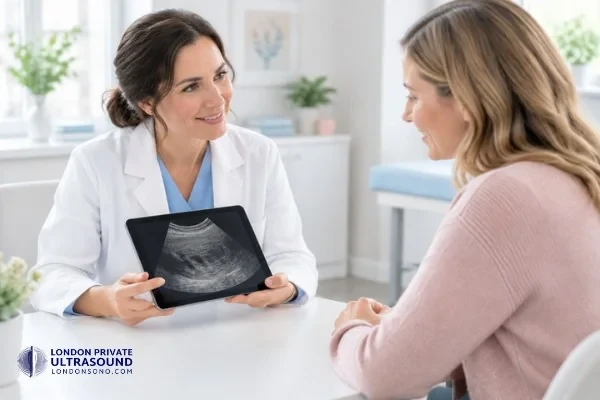
But here’s where an ultrasound really helps: a fibroid and cyst can be differentiated on a scan.
| Feature | Fibroid | Cyst |
|---|---|---|
| What it is | A solid growth of muscle | A fluid-filled sac |
| Where it grows | In or on the womb | Most often on an ovary |
| On an ultrasound scan | A firm, rounded lump of tissue in the wall of the womb | A dark, fluid-filled bubble (simple cyst) |
What Are the Types of Fibroids?
Fibroids are grouped by where they grow in the womb, and doctors use the FIGO classification to describe them in more detail, from type 0 to type 8.4
If you happen to see one of these terms on your report, it’s telling you where the fibroid sits, which shapes the symptoms you’re likely to notice and the treatments that might suit you. Here’s a quick overview:
Submucosal fibroids
Submucosal fibroids grow just under the lining of the womb and bulge into the cavity. Even small ones can cause heavy bleeding, and they’re the type most likely to affect fertility.
Intramural fibroids
Intramural fibroids grow within the muscle wall of the womb. They’re the most common type, and larger ones can make the womb feel bulky and cause heavy periods or pressure.
Subserosal fibroids
Subserosal fibroids grow on the outer surface of the womb. They’re less likely to affect bleeding but can press on your bladder or bowel as they grow.
Pedunculated fibroids
Pedunculated fibroids hang from the womb on a small stalk, either inside the cavity (type 0) or outside it (type 7). Occasionally the stalk can twist, which causes sudden, severe pain and needs urgent help.
How Are Fibroids Diagnosed?
The first step to diagnosing fibroids usually starts with an imaging test, such as an ultrasound scan. Such scans can show how many there are, how big they are and exactly where they sit.
Your GP will usually ask about your symptoms and examine you first, and if you go the NHS route, you’ll be referred for an ultrasound if fibroids are suspected.
But if you’re choosing private healthcare, you can opt for an ultrasound without a GP referral, with same-day appointments, similar to how we offer at London Private Ultrasound.
Ultrasound: the first step
An ultrasound scan is the first step in diagnosing fibroids. A transabdominal scan looks through your lower abdomen with a comfortably full bladder, and a transvaginal scan, which is only ever done with your consent, gives a closer, clearer view of the womb.
In a 2002 study of 106 women, transvaginal ultrasound picked up fibroids in 99% of the women who had them, matching MRI.3
What do fibroids look like on an ultrasound scan?
On ultrasound, fibroids usually look like firm, rounded lumps with clear edges, often with a slightly swirled or striped pattern inside.
Your sonographer measures each one and notes where it sits in relation to the lining and the outer wall of the womb.
Can a CT or MRI scan detect fibroids?
A CT scan can sometimes show large or calcified fibroids, but it isn’t used to diagnose them. This is because ultrasound is a much better option to look at the uterus, without any radiation.
An MRI scan can map fibroids very precisely and is sometimes used before surgery or embolisation.
How Are Fibroids Treated and What Are the Options?
Fibroid treatment is often a personalised plan depending on your symptoms and the size, number and position of your fibroids. Fibroids that aren’t causing symptoms often don’t need treatment, but where symptoms are apparent, or if fibroids may affect fertility, treatment is considered.1,5
Here are the common treatment options:
Medicines
- Painkillers such as ibuprofen can help with painful periods
- Tranexamic acid or hormone treatments, such as the combined pill or a hormonal coil, can reduce heavy bleeding
Less invasive procedures
- Uterine artery embolisation (UAE) blocks the blood supply to the fibroids so they shrink, without major surgery
- Endometrial ablation removes the lining of the womb, which may help with heavy bleeding in some women
Surgery
- A myomectomy removes fibroids while keeping the womb intact, by keyhole (laparoscopic) surgery, open surgery or through the cervix
- A hysterectomy removes the womb so fibroids can’t come back. It’s usually only considered when other treatments haven’t worked and you don’t plan to have children
Some treatments can affect your fertility or future pregnancies, so it’s worth talking openly with your doctor about your plans before you decide. You deserve time to weigh up your options without feeling rushed.
When Should You See a GP About Fibroids?
It’s recommended to see your GP if you have symptoms of fibroids, if they’re affecting your daily life, work or relationships, or if painkillers aren’t helping. You don’t need to wait until things feel unbearable to ask for help.
How Much Does a Fibroid Ultrasound Scan Cost in London?
A fibroid scan in London usually ranges from £190 to £250, and it depends on what’s included.
At London Private Ultrasound, a fibroid ultrasound costs £219, which includes:
- detection, measurement and mapping of every fibroid, including its FIGO type
- a check of the womb lining, ovaries and surrounding area
- a transabdominal scan and, only where it adds information and with your consent, a transvaginal scan
- your findings explained on the day
- a written report with images within 24 hours, ready to share with your GP or gynaecologist
Book a Fibroid Ultrasound Scan in London
We know it can feel worrying to book a pelvic scan, especially when you’ve been living with heavy periods or pain for a while.
But when you book a scan with us at London Private Ultrasound, you’ll be looked after by a calm, experienced team and get clear answers the same day, from a clinic that has been caring for patients in the Harley Street Medical District since 2012.
Here’s why patients usually book scans with us:
Experienced specialistsYour scan is performed and reported by registered ultrasound consultants in a CQC-registered clinic.
Your comfort comes firstA female sonographer is available on request at no extra cost, and a transvaginal scan is only ever done with your consent.
A full fibroid mapEvery fibroid is recorded by number, size, position and FIGO type, which is the detail a gynaecologist needs to plan treatment.
Answers before you leaveWe’ll explain what we’ve seen while you’re still with us, and your written report follows within 24 hours.
A clear next stepIf your findings point to a procedure, a referral letter is available on request for gynaecology or fertility services.
Trusted by patientsWe’re rated 4.8 out of 5 on Google, with over 1,300 reviews.
You’ll find us at 27 Welbeck Street, a short walk from Bond Street station, and at 54–56 Victoria Street, St Albans.
Book your fibroid scan or call us on 020 7101 3377. You can also message us on WhatsApp.
Other Scans and Packages for Fibroids and Pelvic Health
Frequently Asked Questions
Can an ultrasound detect all fibroids?
Ultrasound picks up most fibroids, especially with a transvaginal scan. Very small fibroids, or a very large or bulky womb, can be harder to see, and an MRI is occasionally used for a more detailed map.
Do fibroids affect fertility?
Most women with fibroids can get pregnant without problems. Fibroids that bulge into the womb cavity (submucosal fibroids) are the type most likely to affect fertility, which is why knowing their position helps.
Can fibroids turn into cancer?
Fibroids are non-cancerous. Very rarely, a fast-growing lump in the womb turns out to be a different, cancerous growth, so let your doctor know if a fibroid grows quickly, especially after menopause.
Can I shrink fibroids naturally?
There’s no proven way to shrink fibroids with diet or supplements. Fibroids often shrink after menopause, and medical treatments can reduce their size or ease symptoms in the meantime.
Do I need a GP referral for a fibroid scan?
No, when you’re going ahead with private healthcare, you don’t need a GP referral for your scans. At London Private Ultrasound, you can book directly with us and share your report with your GP or gynaecologist afterwards.
Medical Reviewer

Private GP · MBBS BSc (Hons) MRCGP
Trained at Imperial College London and a Member of the Royal College of General Practitioners.
Published Last reviewed
References
- NHS. Fibroids. www.nhs.uk/conditions/fibroids/
- Baird DD, Dunson DB, Hill MC, Cousins D, Schectman JM. High cumulative incidence of uterine leiomyoma in black and white women: ultrasound evidence. Am J Obstet Gynecol. 2003;188(1):100–107. pubmed.ncbi.nlm.nih.gov/12548202/
- Dueholm M, Lundorf E, Hansen ES, Ledertoug S, Olesen F. Accuracy of magnetic resonance imaging and transvaginal ultrasonography in the diagnosis, mapping, and measurement of uterine myomas. Am J Obstet Gynecol. 2002;186(3):409–415. pubmed.ncbi.nlm.nih.gov/11904599/
- Munro MG, Critchley HOD, Fraser IS; FIGO Menstrual Disorders Committee. The two FIGO systems for normal and abnormal uterine bleeding symptoms and classification of causes of abnormal uterine bleeding in the reproductive years: 2018 revisions. Int J Gynaecol Obstet. 2018;143(3):393–408. pubmed.ncbi.nlm.nih.gov/30198563/
- NICE. Heavy menstrual bleeding: assessment and management (NG88). www.nice.org.uk/guidance/ng88