
1. Thyroid & Neck
Thyroid gland, salivary glands, neck lymph nodes, and any palpable neck lumps or bumps. Can help identify nodules, goitre, thyroiditis, and lymphadenopathy.
The Full Body Ultrasound is a comprehensive ultrasound screening covering major organs from neck to pelvis in a single visit. £600 all-inclusive. No GP referral required.
A Full Body Ultrasound Scan at London Private Ultrasound is a comprehensive 40-minute examination covering major body organs — including the carotid arteries, thyroid and neck, liver, pancreas, spleen, gallbladder, kidneys and bladder, uterus and ovaries for women / prostate and testes for men. Performed by an experienced consultant sonographer or registered healthcare professional specialising in ultrasound imaging. From £600 all-inclusive. Same-day verbal results and written report within 24 hours. No GP referral required.
A thorough preventive screening in a single appointment. Six comprehensive ultrasound examinations performed back-to-back, with same-day verbal findings and one integrated written report. Same-Day Results & Tailored Advice.
| Price | £600 all-inclusive · no hidden fees |
|---|---|
| What’s included | Carotid Arteries, Thyroid and Neck, Liver, Pancreas, Spleen, Gallbladder, Kidneys and Bladder, Uterus and Ovaries for Women / Prostate and Testes for Men |
| Duration | ~40–60 minutes total |
| Who performs it | Consultant Sonographer or Vascular Scientist (specialist for each body region) |
| Best for | Comprehensive body screening · health-conscious adults · pre-illness baseline · annual MOT · anxiety about general health |
| Same-day appointments | Yes — typically available, including weekends |
| GP referral required? | No — direct booking |
| Locations | Central London (27 Welbeck Street) and St Albans |
| Results | Same-day verbal scan results · Written report within 24 hours |
Your Full Body Ultrasound covers the most important body systems that can be assessed non-invasively with ultrasound in a single 40-minute appointment. Each examination is performed using the same imaging protocols and clinical standards as an individual standalone scan, with findings explained in clinical context.
Thyroid gland, salivary glands, neck lymph nodes, and any palpable neck lumps or bumps. Can help identify nodules, goitre, thyroiditis, and lymphadenopathy.
Both carotid arteries assessed for plaque and narrowing (stenosis), which may contribute to stroke risk. Doppler flow analysis helps identify clinically significant narrowing affecting blood flow.
Liver, gallbladder, pancreas, spleen, and abdominal aorta. Detects fatty liver, gallstones, masses, and abdominal aortic aneurysm (AAA).
Both kidneys and bladder. Detects kidney stones, cysts, masses, hydronephrosis, and bladder abnormalities.
Comprehensive assessment of accessible lymph node groups. Identifies enlarged or abnormal nodes that may indicate infection, inflammation, or other conditions.
Booking these six ultrasound scans separately would mean six different appointments, six separate reports, six different visits to our clinic, and over £1,344 in fees. By performing all six examinations back-to-back in a single 40-minute appointment, we deliver substantial efficiency. We pass the savings to you.
| Thyroid & Neck Ultrasound | £219 |
| Carotid Doppler | £249 |
| Abdominal Ultrasound | £219 |
| Kidneys & Urinary Tract | £219 |
| Pelvis / Testes | £219 |
| Lymph Node Ultrasound | £219 |
| Total when booked separately | £1,344 |
| Full Body Ultrasound package | £600 |
| You save | £744 |
Beyond the cost saving, you also receive: one integrated report covering all findings in clinical context (rather than six separate reports), a single visit instead of six, and same-day verbal results explained by your ultrasound consultant.
Three levels of comprehensive ultrasound screening — choose the depth of assessment that suits your needs. All performed by registered ultrasound consultants in a single visit.
Ultrasound is a powerful diagnostic tool that uses high-frequency sound waves to image internal organs in real time, without ionising radiation. Below are the most clinically significant findings the Full Body Ultrasound can detect across each body region.
A single 40-minute appointment from arrival to written report. Each ultrasound examination is performed by a registered ultrasound consultant, with all findings discussed in clinical context at the end of the visit.
Welcome at reception, brief medical history review, and any specific symptoms you’d like the consultant to focus on. Quick preparation in the consultation room.
Your consultant begins with the upper body — thyroid gland, salivary glands, neck lymph nodes, and Doppler assessment of both carotid arteries.
Comprehensive abdominal ultrasound: liver, gallbladder, pancreas, spleen, and abdominal aorta. Each organ assessed systematically with images saved for the report.
Both kidneys and bladder assessed. For optimal bladder imaging, a full bladder is required — hence the pre-appointment water intake instruction.
For women: pelvic ultrasound covering uterus, ovaries, endometrium, and adnexa. For men: testicular and prostate examination. Lymph node assessment completes the scan.
Your consultant explains all findings on the day, answers your questions, and outlines any recommendations. A comprehensive written report with images is delivered within 24 hours.
Few private clinics in London offer this breadth of ultrasound assessment within a single appointment at this price point. Performed by registered ultrasound consultants under the supervision of a GMC-registered Medical Director.
Every Full Body Ultrasound is performed by a registered ultrasound consultant or qualified sonographer — never delegated. Quality is non-negotiable.
Latest-generation ultrasound systems with high-frequency linear and curved array transducers — the same equipment used in leading NHS teaching hospitals.
You don’t wait days for findings. Your consultant explains everything on the day, with the comprehensive written report and saved images delivered within 24 hours.
Six separate reports tell you about six separate organs. Our integrated report tells you what your overall picture means — findings in clinical context, with onward guidance where needed.
Fully registered with the Care Quality Commission. Medical oversight by a GMC-registered Medical Director. CQC Champion ensures ongoing compliance.
Available at our flagship clinic at 27 Welbeck Street in the Harley Street medical district, and at our St Albans clinic. Both offer the full Full Body Ultrasound service.
Everything you need to know about the Full Body Ultrasound Scan at London Private Ultrasound.
A Full Body Ultrasound Scan is a comprehensive 60-minute examination covering 6 major body regions in a single appointment:
Performed by a registered ultrasound consultant using premium imaging equipment. From £600 all-inclusive. No GP referral required.
Full Body Ultrasound Scan costs £600 all-inclusive. The fee includes:
When booked individually, the same six scans would cost over £1,344. The package represents savings of more than £700.
The Full Body Ultrasound includes six comprehensive examinations:
When you book six ultrasound scans separately:
By performing all six examinations back-to-back in a single 40-minute appointment, we deliver substantial efficiency. We pass these savings to you: £600 instead of £1,344+. You also receive:
Your Full Body Ultrasound is performed by one of our registered ultrasound consultants:
All are qualified ultrasound consultants or sonographers registered with relevant UK professional bodies including BMUS (British Medical Ultrasound Society), SVT (Society for Vascular Technology), and the Society of Radiographers. Medical oversight is provided by our Medical Director, Dr Babak Soleimanpour (GMC: 6060555).
The Full Body Ultrasound can detect a wide range of conditions:
Any findings are explained on the day and a written report with onward referral guidance is provided.
For optimal imaging quality:
The scan is non-invasive, painless, and uses no ionising radiation. You can drive yourself to and from the appointment, and return to normal activity immediately afterward.
No. You do not require a GP referral. Self-refer directly online at londonsono.com or call 020 7101 3377.
Same-day appointments are often available, including weekends. Both clinics offer the Full Body Ultrasound:
Many of our patients are concerned that if they come to us, they won’t be able to go back to public health. That simply is not the case. Our goal is to give you a comprehensive screening as quickly as possible and get you on the right pathway — no strings attached.
If your scan identifies a condition requiring NHS specialist management — for example a suspicious lesion, abdominal aortic aneurysm requiring vascular surgery follow-up, or a finding that needs onward investigation — we generate a same-day formal referral letter for your GP at no additional charge.
The referral is significantly stronger because it includes confirmed imaging findings — meaning your specialist appointment is more productive from the first visit.
The three tiers cover progressively more comprehensive screening:
Our team are fully registered and regulated for practice in the United Kingdom. Our patients should expect nothing less.
Your Full Body Ultrasound is delivered by a multi-disciplinary team of registered ultrasound consultants, sonographers, and supporting clinicians — all working under the medical supervision of our Medical Director.

Clinical Vascular Scientist · BMUS: 20702 · SVT: 679 · SOR: 93290 · Co-founder of London Private Ultrasound
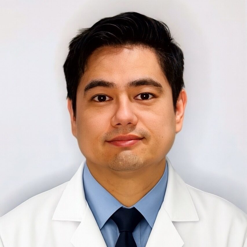
Registered sonographer specialising in general medical and vascular ultrasound
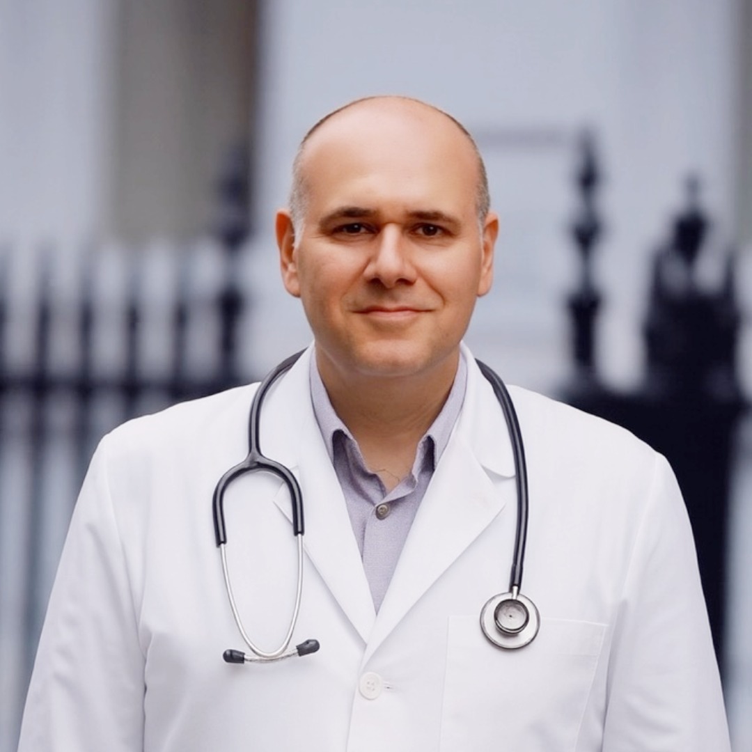
MD · MRCGP · DRCOG · GMC: 6060555 · Medical Director with overall clinical governance responsibility

Co-founder of London Private Ultrasound · CQC Champion ensuring compliance with Care Quality Commission standards
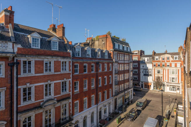
Reassurance and peace of mind are at the heart of everything we do. Located at 27 Welbeck Street in the Harley Street medical district. Full Body Ultrasound also available at our St Albans clinic at 54–56 Victoria Street.


Use the secure booking calendar to select your preferred clinic, date, and time. Same-day appointments often available. For advice on whether Full Body, Plus, or Premium is right for you, call 020 7101 3377.
Secure online booking
Choose a time that suits you — we search every eligible specialist and confirm your appointment instantly.
Select any available day in the next 60 days.
Choosing a specialist re-draws the calendar to show only their available dates.
All fields are required unless marked optional.
Use the secure Stripe tabs below to select Card, Google Pay, Apple Pay or Klarna when available.
Payment methods shown depend on your device and the options enabled in Semble Pay. Card details are handled securely by Stripe and are not stored by this website.
Thank you, — a confirmation email has been sent to .
Your scan is booked. Anything selected below will be attached to the same booking and can be paid for at the clinic.
Three ways to book your Full Body Ultrasound — or to find out whether Full Body, Plus, or Premium is the right tier for your screening goals.
Use our secure booking platform to choose your preferred clinic and date. Instant confirmation by email. Available 24 hours, 7 days a week.
Book Full Body Ultrasound online Available 24 hours, 7 days a weekUnsure which tier is right for you? Want to discuss your specific concerns? Call us — our team will help you choose the right level of screening.
020 7101 3377 Mon–Sun · 9am–7pmHave prior imaging to share or want to discuss complex history before booking? Email our team with the details.
[email protected] We respond within a few hours during clinic hoursLatest health insights, clinic news, and patient stories from London Private Ultrasound.