Musculoskeletal ultrasound · Harley Street, Central London & St Albans · 7 days a week

Private Hip Ultrasound Scan in London

A private hip ultrasound scan at London Private Ultrasound costs £199 for one hip, or £299 for both hips, with no GP referral needed. It is a painless, radiation-free hip and upper thigh scan covering the gluteal tendons and trochanteric bursae on the outer hip, the iliopsoas tendon and bursa and hip flexors at the front, the groin and adductors, the hamstring origin at the sit bone, the front of the hip joint and the muscles of the upper thigh. It diagnoses greater trochanteric pain syndrome, trochanteric bursitis, gluteal tendon tears, iliopsoas bursitis, hip flexor and groin strains, snapping hip, thigh muscle tears, haematoma and soft-tissue lumps. It takes about 25–35 minutes, includes a dynamic assessment that MRI cannot provide, and comes with same-day results and a written report within 24 hours.

A deep ache on the bony point of the hip that is worse lying on that side at night, groin pain that bites when you put your socks on, a hamstring that has never been right since it went, a click or snap deep in the hip, or a lump in the thigh you cannot explain: a hip ultrasound is the recommended first-line scan for most soft-tissue hip and thigh problems. You are seen by an MSK-specialist clinician who scans the hip in real time, presses on the exact spot that hurts to match the pain to the picture, explains the findings in the room, and sends a full written report.

  • £299 both hips · £199 one hip
  • Hip and upper thigh included
  • Gluteal tendons & trochanteric bursa
  • Groin, hip flexors & hamstring origin
  • Snapping hip seen in motion
  • MSK-specialist clinicians
  • Same-day results · report in 24 hours
  • No GP referral needed
Why most people book both hips

A single-hip scan covers the outer hip, groin, buttock and upper thigh on the symptomatic side. Scanning both hips is the better examination clinically: gluteal tendon thickness and bursal fluid vary from person to person, so the most reliable way to judge your painful side is to measure it against the other one, and it catches the early change on the quiet hip that is so common in greater trochanteric pain syndrome. Your scan fee covers the appointment, the findings explained on the day and the written report. If what we find calls for more — a GP or consultant consultation, an ultrasound-guided injection or an onward referral — we can arrange it here, usually quickly, and it is booked and charged separately.

Quick facts

Hip Ultrasound Scan at a Glance: Cost, What It Shows and Where

Price£299 both hips (saving £99) · £199 one hip · all-inclusive: scan, results & written report
Also known asHip scan · hip and upper thigh scan · hip and thigh ultrasound · MSK hip ultrasound · gluteal tendon scan
What is assessedGluteus medius & minimus tendon insertions, greater trochanteric bursae, iliotibial band, iliopsoas tendon & bursa, hip flexors & rectus femoris origin, adductor origins & groin, femoral canal, anterior hip joint recess, hamstring origin at the ischial tuberosity, upper thigh muscles, lateral femoral cutaneous nerve & soft-tissue lumps
DiagnosesGreater trochanteric pain syndrome, trochanteric bursitis, gluteal tendinopathy & tears, calcific tendinopathy, iliopsoas bursitis, hip flexor & groin strain, snapping hip, hip joint effusion & synovitis, proximal hamstring tendinopathy & tears, thigh muscle tears & haematoma, meralgia paraesthetica, hernias, lumps & cysts
Appointment timeAbout 25–35 minutes per hip
PreparationNone. Wear or bring loose shorts so the outer hip, groin and upper thigh can be reached. No fasting, no radiation
GP referralNot required · book directly. Physiotherapist, osteopath and consultant referrals welcome
ResultsExplained on the day · written report within 24 hours for your GP, physio or specialist
TreatmentUltrasound-guided hip or bursa injection arranged where appropriate
Locations27 Welbeck Street, Harley Street, London W1G 8EN · 54–56 Victoria Street, St Albans AL1 3HZ
Availability7 days a week, 9am–7pm · same-day & next-day hip appointments often available
Two ways to book

Hip and Upper Thigh Ultrasound: Which Option Should You Go For?

Both hips is the option most patients choose, and for hip pain it is usually the better scan rather than just the cheaper pair. Gluteal tendon thickness, bursal fluid and hamstring origin appearances vary a great deal between individuals, so the clearest way to judge whether your painful side is genuinely abnormal is to measure it against your other side in the same sitting. Both options are performed by the same MSK-specialist clinicians, with the same dynamic assessment, same-day results and written report.

Most booked · best value

Hip Scan: Both Hips

Both sides · comparison included

£299

The recommended option. Everything in the single-hip scan on both sides, with a direct left-versus-right comparison of the gluteal tendons and bursae, in one visit with one report. Two separate single-hip appointments would cost £398.

  • Everything in the single-hip scan, both sides
  • Direct left-versus-right tendon comparison
  • Catches early change on the quiet hip
  • Ideal for runners, walkers & hip pain with no injury
  • Inflammatory arthritis & bilateral bursitis review
  • Save £99 · one visit, one report
Book Both Hips · £299

Hip Scan: One Hip

Single side · about 25–35 minutes

£199

The right choice when one hip is clearly symptomatic after a specific injury and you only need that side reported. A full hip and upper thigh assessment on the affected side, with dynamic testing.

  • Gluteal tendons & trochanteric bursae
  • Iliopsoas tendon, bursa & hip flexors
  • Groin, adductors & hernia check
  • Hamstring origin & upper thigh muscles
  • Anterior hip joint & Doppler assessment
  • Same-day results · report in 24 hours
Book One Hip · £199

Not sure which side to book? Hip pain often refers, and the side that hurts is not always the side that is driving it: a painful right hip in someone who limps on the left is a common pattern, as is bilateral gluteal tendinopathy where only one side has become painful yet. If you have symptoms on both sides, if there was no obvious injury, or if physiotherapy has stalled without a clear diagnosis, book both hips. Call 020 7101 3377 if you would rather talk it through first.

What is included

What a Hip and Upper Thigh Ultrasound Scan Includes

A hip ultrasound, sometimes advertised as a hip and upper thigh scan or simply a hip scan, is a systematic examination of the region from four directions: the outside of the hip, the front and groin, the back and buttock, and the thigh itself. Because this MSK hip scan is performed live, the clinician can load the tendons and move the hip while watching them work, which is how snapping, tendon tears and subtle bursal problems are picked up. The upper thigh is included as standard at no extra cost.

Gluteal tendons & the outer hip

The gluteus medius and minimus insertions onto the greater trochanter are measured and assessed for thickening, degeneration, calcification and partial or full-thickness tears — the true cause of most pain on the bony point of the hip. Doppler shows how active the process is.

Trochanteric bursae & iliotibial band

The bursae that sit between the gluteal tendons and the bone are checked for fluid and thickening, and the iliotibial band is followed over the trochanter and tested in movement, because an external snapping hip is generated exactly here.

Iliopsoas tendon, bursa & hip flexors

At the front, the iliopsoas tendon and its bursa are assessed for iliopsoas bursitis and tendinopathy and watched dynamically for an internal snap, along with the rectus femoris and sartorius origins where hip flexor strains occur.

Groin, adductors & hernia check

The adductor origins and the pubic region are examined for groin strain and tendinopathy, and the inguinal and femoral canals are checked, with a cough or strain manoeuvre, for an inguinal or femoral hernia — a frequent and easily missed cause of groin pain.

Hip joint recess & hamstring origin

The anterior hip joint recess is assessed for effusion and active synovitis on Doppler, and at the back the hamstring origin at the sit bone is examined for proximal hamstring tendinopathy, tears and ischiogluteal bursitis — the classic cause of deep buttock pain on sitting.

Upper thigh, nerves & your report

The quadriceps, adductor and hamstring muscles of the upper thigh are scanned for tears, haematoma and scarring, the lateral femoral cutaneous nerve is checked where meralgia paraesthetica is suspected, and any lump is characterised. Findings are explained on the day, with a full written report and images within 24 hours.

What the scan finds

What Can a Hip Ultrasound Scan Detect?

Hip pain has many possible sources, and the job of the scan is to pin down which structure is responsible. An ultrasound for hip pain identifies the specific structural cause — from the gluteal tendons and trochanteric bursa to the iliopsoas, the hamstring origin and the thigh muscles — so you know what you are dealing with and what to do next. These are the conditions a hip and upper thigh ultrasound detects most often.

Greater trochanteric pain syndrome

The umbrella diagnosis for pain on the bony point of the outer hip: a deep ache, worse lying on that side at night, worse on stairs and worse standing on one leg. Ultrasound separates the tendon component from the bursal component, which is what decides whether the answer is loading or an injection.

Gluteal tendinopathy & tendon tears

The gluteus medius and minimus tendons are the hip’s equivalent of a rotator cuff, and they are the primary problem in most outer hip pain. The scan shows thickening, disorganisation, calcific deposits and partial or full-thickness tears, and grades them against the other side.

Trochanteric bursitis & hip bursa fluid

Fluid and thickening in the hip bursa over the greater trochanter, measured directly. Imaging has shown bursal swelling is usually secondary to the tendon problem rather than the cause, which is why treating it as pure inflammation with rest alone so often disappoints. Where it is genuinely the driver, it responds well to a guided injection.

Iliopsoas bursitis & hip flexor strain

Pain and catching at the front of the hip and groin. The iliopsoas bursa can distend, sometimes communicating with the joint, and the tendon can become thickened and painful. Hip flexor and rectus femoris strains are shown directly, with the grade and the extent of any tear.

Snapping hip, internal & external

A snap or clunk you can feel and often hear. Ultrasound is the test of choice because it is dynamic: the iliopsoas flicking over the front of the pelvis, or the iliotibial band and gluteus maximus jumping over the trochanter, are seen live as they happen. A static MRI can miss this entirely.

Groin strain, adductor injury & hernia

Adductor origin tendinopathy and tears, pubic-region pain in footballers and runners, and inguinal or femoral hernias assessed with a cough or strain manoeuvre. Groin pain has several very different causes and this is where a scan earns its place.

Proximal hamstring problems

Deep buttock and sit-bone pain that is worst on sitting, driving or lengthening the leg. The hamstring origin is examined for tendinopathy, partial and complete tears and avulsion, and for ischiogluteal bursitis, which is a common cause of the same pain and is treated differently.

Thigh muscle tears & haematoma

Quadriceps, hamstring and adductor tears are graded and measured, and a haematoma or a dead leg is followed to see whether it is resolving, organising or forming a collection. Serial scanning is used to guide a safe return to sport.

Hip effusion, synovitis & gout

Fluid and active inflammation in the front of the hip joint from inflammatory arthritis, gout or after trauma, with Doppler to show how active it is. Where appropriate, the joint or bursa can be aspirated or injected under live ultrasound guidance.

Meralgia paraesthetica & nerve pain

Burning, tingling or numbness over the outer thigh from compression of the lateral femoral cutaneous nerve, often after weight change, pregnancy, a tight belt or surgery. The nerve is imaged directly and followed to the point where it is being compressed.

Lumps, cysts & collections

A lump on the hip, buttock or thigh is measured and characterised: which layer it sits in, whether it is solid or fluid-filled, and whether it has a blood supply. That distinguishes a lipoma, ganglion, sebaceous cyst, bursa, seroma or old haematoma from something needing further assessment.

After a hip replacement

Ultrasound assesses the soft tissues around a hip replacement where X-ray is weakest: fluid and periprosthetic collections, abductor tendon integrity after surgery, iliopsoas irritation against the cup, and soft-tissue masses. Implant loosening and wear remain X-ray and CT questions.

Being straight about what ultrasound sees at the hip. Ultrasound is excellent for everything around the joint — the gluteal tendons, the bursae, the hip flexors and groin, the hamstring origin, the thigh muscles, the nerves and any lumps — and it is the only common test that shows the hip moving and under load. The inside of the joint is a different question: the hip sits deep, so the labrum, the cartilage, the bone and the shape changes of impingement need an X-ray or MRI. If your symptoms point that way, we will tell you on the day and advise on the right next step.

Seek urgent medical care straight away if you cannot bear weight after a fall or if the leg looks shortened or turned outwards (this needs an X-ray to exclude a hip fracture), if the hip is hot, red and swollen with fever or you feel unwell, if you have a hip replacement and develop increasing pain, fever or a discharging wound, if you felt a sudden tearing at the sit bone with severe bruising down the back of the thigh, or if hip and thigh swelling comes with a painful, tight, swollen calf, which can signal a clot. A child who is limping with a fever or will not weight-bear needs same-day assessment. Contact your GP, NHS 111, or attend urgent care or A&E as appropriate.

The other half of the scan

Hip and Upper Thigh Scan: What the Thigh Part Covers

A hip scan and a hip and upper thigh scan are the same appointment described two different ways, and at London Private Ultrasound the thigh is included as standard. It matters more than it sounds, because a great deal of what patients call hip pain actually arises in the upper thigh: an adductor strain in the groin, a torn quadriceps, a hamstring origin that never healed properly, a dead leg that has organised into a collection, or a lump in the thigh that needs characterising. If your scan stops at the outer hip, those causes are simply not looked at.

Quadriceps & rectus femoris

The quadriceps group is scanned along the front of the thigh for muscle tears, the classic dead-leg contusion and haematoma, and for the rectus femoris origin at the front of the pelvis, a frequent site of injury in footballers, sprinters and kickers.

Adductors & groin

The adductor longus origin is the most commonly injured structure in groin pain. Tendinopathy, partial tears and complete avulsions are shown directly, and the surrounding pubic region is assessed where groin pain has become chronic.

Hamstrings, from origin to mid-thigh

The hamstring origin at the sit bone and the muscle bellies down the back of the thigh are assessed for tendinopathy, partial and complete tears, retraction and old scarring, which is what determines rehabilitation timelines and return-to-sport decisions.

Haematoma, seroma & collections

Bleeding within or between muscles after an injury, injection, fall or surgery is measured and followed. Ultrasound distinguishes a resolving bruise from an organising collection that may need draining, and can guide aspiration where appropriate.

Thigh lumps & masses

Any lump in the thigh is measured and characterised by depth, layer, internal structure and blood flow. Most are straightforward — lipomas, cysts, ganglions, muscle hernias — and where anything looks indeterminate we say so and arrange the right next step.

Nerves & vessels of the thigh

The lateral femoral cutaneous nerve is assessed in suspected meralgia paraesthetica, and the femoral vessels are checked for a pseudoaneurysm or collection after catheterisation or injection. A suspected clot in the leg veins is a separate DVT Doppler scan.

Where the hip and thigh scan stops. This appointment covers the hip, buttock, groin and thigh down towards the knee. Pain at the knee itself is a knee ultrasound, and calf pain is a calf scan or a DVT Doppler depending on the symptoms. Buttock pain that shoots down the back of the leg with pins and needles is usually referred from the lower back rather than the hip, and is better assessed clinically and with an MRI of the spine. If you are unsure, call 020 7101 3377 and we will tell you which appointment to book before you pay for one.

Your appointment

How a Hip Ultrasound Is Done, and How to Prepare

There is nothing to do beforehand: no fasting, no medication changes, no radiation. The scan is gentle and painless, and you can walk, drive and return to your day straight afterwards. The one thing that helps is clothing you can move out of the way, because the outer hip, the groin and the upper thigh all need to be reached.

When you bookTell us

Say exactly where it hurts

Point us to the area: the bony point on the outside of the hip, the groin or front, the sit bone and buttock, or the thigh. Tell us whether it is one hip or both, how it started, and mention any previous scans, injections, hip replacement or surgery. Choose the both-hip scan if both sides are symptomatic, or if there was no clear injury.

On the dayWear

Wear or bring loose shorts

Loose shorts are ideal, and worth bringing if you are coming from work. Gowns and towels are provided, only the area being scanned is uncovered, and a female clinician can be requested at booking, subject to availability.

BringReports

Bring any previous imaging

If you have earlier hip X-ray, ultrasound or MRI reports, bring them so the clinician can read today’s findings in context. If a physiotherapist, osteopath or consultant sent you, bring their letter and we will report back to them.

DuringAssess

Expect to change position and move the hip

Most of the scan is done lying on your side, which brings the gluteal tendons and bursae directly under the probe. You then lie on your back for the groin, hip flexors and joint, and face down or side-lying for the hamstring origin. You will be asked to move the hip and lift or roll the leg so the tendons can be seen under load and any snapping reproduced. Pressing on the tender spot is deliberate: it matches your pain to what is on the screen.

AfterResults

Findings explained before you leave

The clinician talks you through what they have found on the screen, what it means and what the sensible next step is. A full written report with images follows the same day or within 24 hours, written so your GP, physiotherapist or surgeon can act on it. If treatment is appropriate, a guided injection can often be arranged.

Why us

Why People Choose London Private Ultrasound for Their Hip Scan

Hip scans are widely advertised and prices vary. What matters underneath is who performs and reports the scan, whether the upper thigh and groin are actually included, whether the assessment is dynamic, how quickly you get your report, and whether treatment can follow in the same place. These are the things worth comparing before you book anywhere.

MSK-specialist clinicians

Your hip is scanned and reported by clinicians with specialist musculoskeletal expertise, a consultant-led team combining orthopaedic surgery, MSK ultrasound and radiology. Every scan is personally interpreted by the clinician who performs it, not sent to a remote reader.

The whole region, not just the outer hip

The groin, the hamstring origin and the upper thigh are included as standard, because hip pain frequently comes from there. Some clinics scan the trochanter alone; if your problem is an adductor strain or a hamstring origin tear, that scan will come back normal.

Dynamic, real-time assessment

We move and load the hip while scanning, so a snapping iliopsoas or iliotibial band is captured as it happens and tendons are judged under load. This is the single biggest advantage ultrasound has over a static MRI for hip pain.

Results the same day

Findings are explained in the room, and a full written report with images follows within 24 hours, ready to share with your GP, physiotherapist, osteopath or surgeon.

Harley Street, seven days a week

A CQC-registered clinic at 27 Welbeck Street in the Harley Street Medical District, plus St Albans for Hertfordshire, open 9am to 7pm every day, with no referral needed and same-day appointments often available. Established 2012, rated 4.8 across more than 1,200 reviews.

Book online

Book Your Private Hip and Upper Thigh Ultrasound

£299 for both hips or £199 for one, with your findings explained on the day and a full written report within 24 hours. Both hips is the option most people choose: it saves £99 against two separate visits and gives the clinician your other side to measure against. Same-day and next-day appointments are often available at both clinics, seven days a week. Call 020 7101 3377 if you would like to talk it through first, to request a female clinician, or to arrange a baby or child hip scan.

Secure online booking Choose both hips or one · 7 days a week

Choose your appointment

Open secure booking to view available hip scan appointments. Choose one hip or both hips. For baby or child appointments, call our team to check suitability first.

Related services

The other musculoskeletal appointments our hip patients book most often, at both clinics, seven days a week.

Ultrasound-Guided Hip Injection

If the scan confirms trochanteric bursitis, gluteal tendinopathy, iliopsoas bursitis or joint synovitis, a precision injection or aspiration placed under live ultrasound can often be arranged for faster relief.

Knee Ultrasound Scan

For knee pain, a suspected meniscal or tendon problem or a Baker’s cyst. Often booked alongside a hip scan when a running or walking problem involves the whole limb.

From £199View scan →

Muscle & Sports Injury Ultrasound

Focused imaging of a muscle tear, a groin or hamstring injury or an overuse problem, with dynamic assessment and a clear return-to-activity picture.

From £199View scan →

Hernia Ultrasound Scan

If your pain is in the groin and worse on coughing, straining or lifting, an inguinal or femoral hernia is an important alternative diagnosis and this is the dedicated scan for it.

From £199View scan →

DVT Doppler Scan — Leg

If hip or thigh swelling comes with calf pain, tightness or redness, a clot is an important alternative diagnosis and this is the correct scan. Same-day urgent appointments are usually available.

From £199View scan →

One-Stop Orthopaedic & MSK Clinic

Scan, consultant review and treatment in a single pathway when you want to move from diagnosis to a plan as quickly as possible.

Find us

Hip Ultrasound Near Me: Central London and St Albans

Two clinics, seven days a week, 9am to 7pm, with same-day and next-day hip appointments frequently available. Both run the full hip and upper thigh ultrasound — £299 for both hips, £199 for one — with MSK-specialist clinicians and a written report within 24 hours.

London Private Ultrasound clinic at 27 Welbeck Street in the Harley Street Medical District, Central London

Central London: Harley Street Medical District

27 Welbeck Street, London W1G 8EN

Two minutes from Bond Street (Elizabeth line, Central, Jubilee), five from Oxford Circus, eight from Marylebone and Baker Street. Convenient for patients travelling in from Mayfair, Fitzrovia, Marylebone, Soho, the City and Paddington, and for anyone searching for a hip ultrasound in London, a hip scan near me, or a hip ultrasound Harley Street clinic. Because Bond Street sits on the Elizabeth line, it is also a direct run from Slough, Reading and the Thames Valley to the west, and from Stratford and Essex to the east.

020 7101 3377 · Get directions

London Private Ultrasound clinic, St Albans, Victoria Street

St Albans: Hertfordshire

54–56 Victoria Street, St Albans AL1 3HZ

A short walk from St Albans City station, with parking nearby. Convenient for patients across Hertfordshire and North London, including Watford, Hatfield, Harpenden, Radlett and Welwyn Garden City.

Contact the clinic · Get directions

Accredited · Regulated · Trusted

Registered & Regulated

Your hip scan sits within a CQC-registered pathway, performed and reported by clinicians on the UK’s professional registers.

The Royal College of Radiologists

Professional standards, education and guidance for clinical radiology and clinical oncology.

General Medical Council

Official register and professional standards for doctors and regulated medical associates.

Care Quality Commission

Independent regulator of health and adult social-care services in England.

Health and Care Professions Council

Official register for regulated health and care professionals, including radiographers.

Royal College of Surgeons of England

Professional standards, education and support for surgical practice and patient care.

4.8Google Rating
1,200+Verified Reviews
35,000+Patients Served
2012Harley Street Est.
Who you will see

Meet the MSK Team Behind Your Hip Scan

A consultant-led musculoskeletal team combining orthopaedic surgery, specialist MSK ultrasound, and musculoskeletal radiology. The clinician who scans you explains your findings, and can arrange treatment where it is needed.

Mr Peyman Bakhshayesh, Consultant Trauma and Orthopaedic Surgeon at London Private Ultrasound

Mr Peyman Bakhshayesh

Principal Consultant · Trauma & Orthopaedic Surgeon

MD · PhD · MBA · multi-joint specialist · ultrasound-guided procedures, PRP & regenerative medicine

Mr Paul Watson, Lead MSK Consultant Clinician at London Private Ultrasound

Mr Paul Watson

Lead MSK Consultant Clinician

NMP · DipMSK · DFSEM · CSP · HCPC · MSK ultrasound & ultrasound-guided injection therapy

Mr Pedram M. Aghaei, Ultrasound Consultant at London Private Ultrasound

Mr Pedram M. Aghaei

Co-founder & Ultrasound Consultant

MD · Clinical Vascular Scientist · BMUS · SoR · MSK and Doppler imaging

Dr Reza Salehi, Musculoskeletal Radiology Doctor at London Private Ultrasound

Dr Reza Salehi

Musculoskeletal Radiology

MBChB · FRANZCR · MSK radiology reporting · extensive private-practice experience

Dr Adil Naeem, Musculoskeletal Radiology Doctor at London Private Ultrasound

Dr Adil Naeem

Musculoskeletal Radiology

MBChB · FRANZCR · MSK radiology reporting · extensive private-practice experience

Dr Babak Soleimanpour, Medical Director and GP at London Private Ultrasound

Dr Babak Soleimanpour

Medical Director & GP

MD · MRCGP · DRCOG · GMC 6060555 · overall clinical governance

Questions

Hip Ultrasound Scan: Frequently Asked Questions

Written and reviewed by our MSK clinical team, answering the questions we are asked most often about hip and upper thigh scans.

How much does a hip ultrasound scan cost in London?

At London Private Ultrasound a hip and upper thigh ultrasound scan is £199 for one hip and £299 for both hips, all-inclusive, at our Central London (Harley Street Medical District) and St Albans clinics. The price covers an MSK-specialist appointment, the full hip and upper thigh assessment, a dynamic assessment, same-day verbal results and a written report with images within 24 hours. There are no added fees, and we will share the report with your GP, physiotherapist or specialist on request.

Across London, private hip ultrasound cost generally runs from around £160 to £250 for a single hip — a hip scan in London is rarely cheaper than that once the report is included — and the difference usually comes down to who performs and reports the scan, how long the appointment is, whether the groin and upper thigh are included or only the outer hip, and whether treatment can follow in the same place. When comparing hip scan cost between clinics, that is what the gap between a £160 scan and a £250 one usually represents. Booking both hips together is £299 rather than £398, a £99 saving against two separate single-hip appointments.

How much does an ultrasound cost in London?

Private ultrasound scans in London generally run from about £100 to £350 for a single area, depending on the region scanned, who performs and reports it, and whether a specialist consultation is included. Musculoskeletal scans such as a hip ultrasound usually sit in the £160 to £300 band, while more complex or multi-region studies cost more.

Ours is £199 for one hip and £299 for both hips, all-inclusive. When comparing quotes, check three things: whether the clinician’s assessment and the written report are included in the headline price, whether an MSK specialist performs and interprets the scan, and how much of the region is actually covered. A cheaper scan that looks only at the trochanteric bursa will not answer a groin or hamstring problem.

What can a hip ultrasound detect?

A hip ultrasound detects the soft-tissue causes of hip, groin and upper thigh pain. That includes greater trochanteric pain syndrome, gluteal tendinopathy and gluteus medius or minimus tendon tears, trochanteric bursitis, calcific tendinopathy, iliopsoas bursitis and hip flexor strain, snapping hip, fluid and active synovitis in the front of the hip joint, adductor and groin strains, proximal hamstring tendinopathy and tears at the sit bone, quadriceps and thigh muscle tears, haematoma and seroma, meralgia paraesthetica from compression of the lateral femoral cutaneous nerve, inguinal and femoral hernias, and soft-tissue lumps such as lipomas, ganglions and cysts.

Because the scan is dynamic, it also shows problems that only appear on movement, such as a snapping iliopsoas or iliotibial band. What it does not assess is the inside of the joint: the labrum, the cartilage, the bone and the shape changes of hip impingement need an X-ray or MRI.

What is a hip and upper thigh ultrasound scan?

A hip and upper thigh ultrasound scan is the same appointment described two ways. Some clinics call it a hip scan, others a hip and upper thigh scan or a hip and thigh ultrasound, and it covers one continuous region: the outer hip and gluteal tendons, the front of the hip and groin, the buttock and hamstring origin, and the muscles of the upper thigh down towards the knee.

At London Private Ultrasound the upper thigh is included as standard at no extra cost, because a great deal of what patients call hip pain arises on the thigh side of the region — an adductor strain, a hamstring origin tear, a quadriceps injury, a thigh lump or a haematoma. If your pain sits at the knee or below, that is a different scan and we will tell you which one to book.

How is a hip ultrasound done?

You change into shorts or a gown and the clinician takes a short history first, because where the pain sits decides how the scan is performed. Most of a hip ultrasound is done lying on your side, which brings the gluteal tendon insertions and the trochanteric bursae directly under the probe. You then lie on your back for the groin, the iliopsoas tendon and bursa, the hip flexors and the front of the hip joint, and face down or side-lying for the hamstring origin at the sit bone.

Warm gel is applied and a small probe is moved over the skin while the structures are viewed on screen. You will be asked to move the hip, lift the leg or roll it in and out so the tendons can be assessed under load and any snapping reproduced. It is painless, takes about 25 to 35 minutes for one hip, and there are no needles and nothing internal. Only the area being scanned is uncovered, and a female clinician can be requested at booking.

Can a hip ultrasound detect arthritis or hip impingement?

Not directly, and it is important to be clear about that. Hip osteoarthritis and femoroacetabular impingement (FAI) are diagnosed on X-ray and MRI, because they involve the joint surfaces, the cartilage, the labrum and the shape of the bone, all of which sit too deep for ultrasound to assess properly.

Ultrasound can show indirect signs, such as fluid and active synovitis in the anterior joint recess or a paralabral cyst, and it is very good at answering a different and very useful question: whether your pain is actually coming from the tendons, bursae and muscles outside the joint, which is a common and highly treatable cause. If your history points at the joint — deep groin pain, stiffness, loss of rotation, difficulty with socks and shoes — we will say so on the day and advise on the right next step rather than sell you the wrong scan.

Ultrasound, X-ray or MRI: which scan do I need for hip pain?

It depends on where the pain sits. Ultrasound is the first-line test for pain on the outside of the hip, in the groin muscles and hip flexors, at the buttock and hamstring origin and in the upper thigh, because those structures are superficial and it shows them moving and under load. X-ray is the correct first test for suspected hip arthritis, for the bony shape in impingement, and after significant trauma where a fracture is possible. MRI is best for the labrum and cartilage, avascular necrosis, stress fractures and bone marrow changes, and for surgical planning.

In practice, outer hip pain is an ultrasound question and deep groin pain with stiffness is usually an X-ray question. Call 020 7101 3377 and describe where it hurts, and we will tell you which one you need before you book.

Can an ultrasound diagnose trochanteric bursitis and gluteal tendinopathy?

Yes, and it separates the two, which matters more than it sounds. Pain on the bony point of the outer hip was labelled trochanteric bursitis for years, but imaging has shown that in most cases the gluteus medius and minimus tendon insertions are the primary problem, with any bursal swelling secondary. The umbrella term now used is greater trochanteric pain syndrome.

Ultrasound measures the tendon insertions, shows thickening, degeneration, calcification and partial or full-thickness tears, uses Doppler to judge how active the process is, and measures any bursal fluid. The distinction changes the treatment: a tendon problem responds to a progressive loading programme rather than rest, while a genuinely distended bursa is the one most likely to respond to an ultrasound-guided injection. Scanning both hips makes this more reliable, because tendon thickness is judged against your other side.

Can ultrasound diagnose a snapping hip or hip flexor problem?

Yes, and this is one of the clearest advantages ultrasound has over MRI. Snapping hip only happens on movement, so a static scan can miss it entirely. During the ultrasound the clinician asks you to move the hip while watching live, so an internal snap from the iliopsoas tendon flicking over the front of the pelvis, or an external snap from the iliotibial band or gluteus maximus over the greater trochanter, is seen and recorded as it happens.

The same appointment assesses the iliopsoas tendon and bursa for iliopsoas bursitis and tendinopathy, and the hip flexors and rectus femoris origin for strain and tears, which covers most searches for a hip flexor ultrasound. Where iliopsoas bursitis is confirmed, a guided injection into the bursa is a well-established treatment.

Does the scan include the groin, the hamstring origin and the thigh?

Yes, all three are part of the same appointment. The groin covers the adductor origins, the pubic region and the femoral canal, where adductor strains, inguinal and femoral hernias and femoral vessel problems are assessed. The hamstring origin at the sit bone is scanned for proximal hamstring tendinopathy, partial and complete tears and ischiogluteal bursitis, which is a common cause of deep buttock pain that hurts most on sitting or driving. The upper thigh covers the quadriceps and rectus femoris, the adductors and the hamstring muscles, for tears, haematoma, scarring and any lump.

This is why the scan is often advertised elsewhere as a hip and upper thigh ultrasound. It is one region and one price with us: £199 for one side, £299 for both.

Do you scan a baby’s hips, and why do breech babies have hip scans?

A baby hip scan is a different examination from a hip ultrasound in adults. A hip ultrasound in adults looks at the soft tissues around a joint that sits too deep to see; in a baby the hip is close to the surface and still largely cartilage, so ultrasound looks straight at the joint itself and X-ray cannot. It is used to check for developmental dysplasia of the hip (DDH), where the socket is shallow or the ball sits partly or completely out of it.

Breech babies are scanned because being in the breech position in late pregnancy is one of the strongest risk factors for DDH, along with a family history of childhood hip problems, being a first-born girl and multiple pregnancy. The NHS newborn examination checks every baby’s hips, and babies with those risk factors or an abnormal examination are referred for an ultrasound, usually at around six weeks of age. The scan is most informative between about six weeks and six months, before the bone hardens enough to block the view.

Baby and child hip scans are arranged by telephone rather than through the online calendar, so please call 020 7101 3377 and our team will confirm suitability, timing and the right clinician. If your baby is already under an NHS paediatric or orthopaedic team, continue with that pathway and bring any reports with you.

Can you scan a child’s or teenager’s hip?

Ultrasound is radiation-free, which makes it well suited to children. It is the test of choice for fluid in the hip joint of a limping child, where transient synovitis — sometimes called irritable hip, and often following a viral illness — is the commonest cause. It is also used for sports and growth-plate related hip and groin pain in adolescents, for a soft-tissue lump, and for suspected muscle injury in young athletes and dancers.

A limping child with a fever, severe pain or an inability to weight-bear needs urgent medical assessment the same day rather than a booked scan, because joint infection has to be excluded first. Remember too that hip problems in children are often felt in the knee. Please call us on 020 7101 3377 before booking a child’s hip scan so we can confirm suitability and arrange the right appointment. A parent or guardian stays in the room throughout.

Can ultrasound check a hip replacement?

Ultrasound is useful for the soft tissues around a hip replacement, which is exactly where X-ray is weakest. It shows fluid and periprosthetic collections, abductor and gluteal tendon integrity after surgery, haematoma and seroma, iliopsoas irritation against the cup, and soft-tissue masses such as the pseudotumours associated with some metal-on-metal implants. It can also guide aspiration where infection is a concern.

What it cannot assess is the implant itself: loosening, wear and component position are X-ray and CT questions, and metal blocks the sound beam so the deep joint is not seen. A suspected infected joint replacement — increasing pain with fever, a hot joint or a discharging wound — needs urgent orthopaedic review, not a routine scan. Please tell us at booking that you have a replacement so we allow the right appointment time.

Can an ultrasound show a hip cyst or a lump on the hip?

Yes, and this is one of the things ultrasound does best. For a lump or cyst on the hip, buttock or thigh, the scan shows how big it is, how deep it sits, which tissue layer it is in, whether it is solid or fluid-filled, and whether it has a blood supply on Doppler. That usually distinguishes the common and harmless findings — a lipoma, a sebaceous or epidermoid cyst, a ganglion, a distended bursa, a seroma or an old haematoma — from something that needs further assessment.

If anything looks indeterminate or concerning, we say so on the day and arrange the right onward step, which may be an MRI or a specialist referral. Any lump that is growing, hard, fixed to deeper tissue, larger than a golf ball or painful should always be assessed.

Is a hip ultrasound painful, and how long does it take?

It is painless and non-invasive. Warm gel is applied to the skin and a small probe is moved over the hip, groin and thigh while the clinician views the structures on screen. There are no needles and nothing internal.

Allow about 25 to 35 minutes for one hip, or a little longer for both, since the examination covers the gluteal tendons and bursae, the groin and hip flexors, the joint recess, the hamstring origin and the upper thigh, and includes a dynamic assessment. Pressing on the tender spot can briefly reproduce your symptoms, which is deliberate and helps match the pain to the picture; tell the clinician if a position is uncomfortable and they will adapt. You can walk, drive and carry on with your day straight afterwards.

How long is the wait for an ultrasound on the NHS?

The NHS operational standard is that patients should wait no longer than six weeks for a diagnostic test such as an ultrasound, but waits vary widely by area and by scan type, and published NHS diagnostic waiting-time data has shown a substantial proportion of patients waiting beyond that. For musculoskeletal scans there are usually two further delays on top: getting a GP appointment and referral in the first place, often after a period of physiotherapy, and then waiting for the report to be issued and returned.

A private hip scan is for people who want a fast, specialist answer, frequently the same or next day, with the findings explained in the room and a written report they can take straight to their GP, physiotherapist or surgeon. We work alongside the NHS rather than around it: the report is written so an NHS team can act on it, and where something needs urgent orthopaedic attention we write a referral letter the same day.

Do I need a GP referral for a private hip scan?

No, a GP referral is not required. You can self-refer and book directly using the booking calendar on this page, by phone on 020 7101 3377, or on WhatsApp, with same-day and next-day appointments often available at both clinics.

Referrals from physiotherapists, osteopaths, sports therapists, consultants and GPs are equally welcome, and we report back to them on request. If you have previous hip X-ray, ultrasound or MRI reports, bring them so the clinician can read today’s findings in context.

Should I book one hip or both hips?

Both hips is the option most patients choose and the one we recommend for most people. It is £299 rather than £398 for two separate single-hip appointments, a £99 saving, and it is done in one visit with one clinician and one report covering both sides.

The clinical reason matters more than the price. Gluteal tendon thickness and bursal fluid vary considerably from person to person, so the most reliable way to judge whether your painful side is genuinely abnormal is to measure it against your other side. Bilateral scanning also catches early change on the quiet hip, which is common in greater trochanteric pain syndrome, and it is the sensible choice for runners and walkers, for anyone being assessed for inflammatory arthritis, and for hip pain with no clear injury. A single hip at £199 is the right choice when one side is clearly symptomatic after a specific injury and you only want that side reported.

Can the scan guide a hip injection or an iliopsoas bursitis treatment?

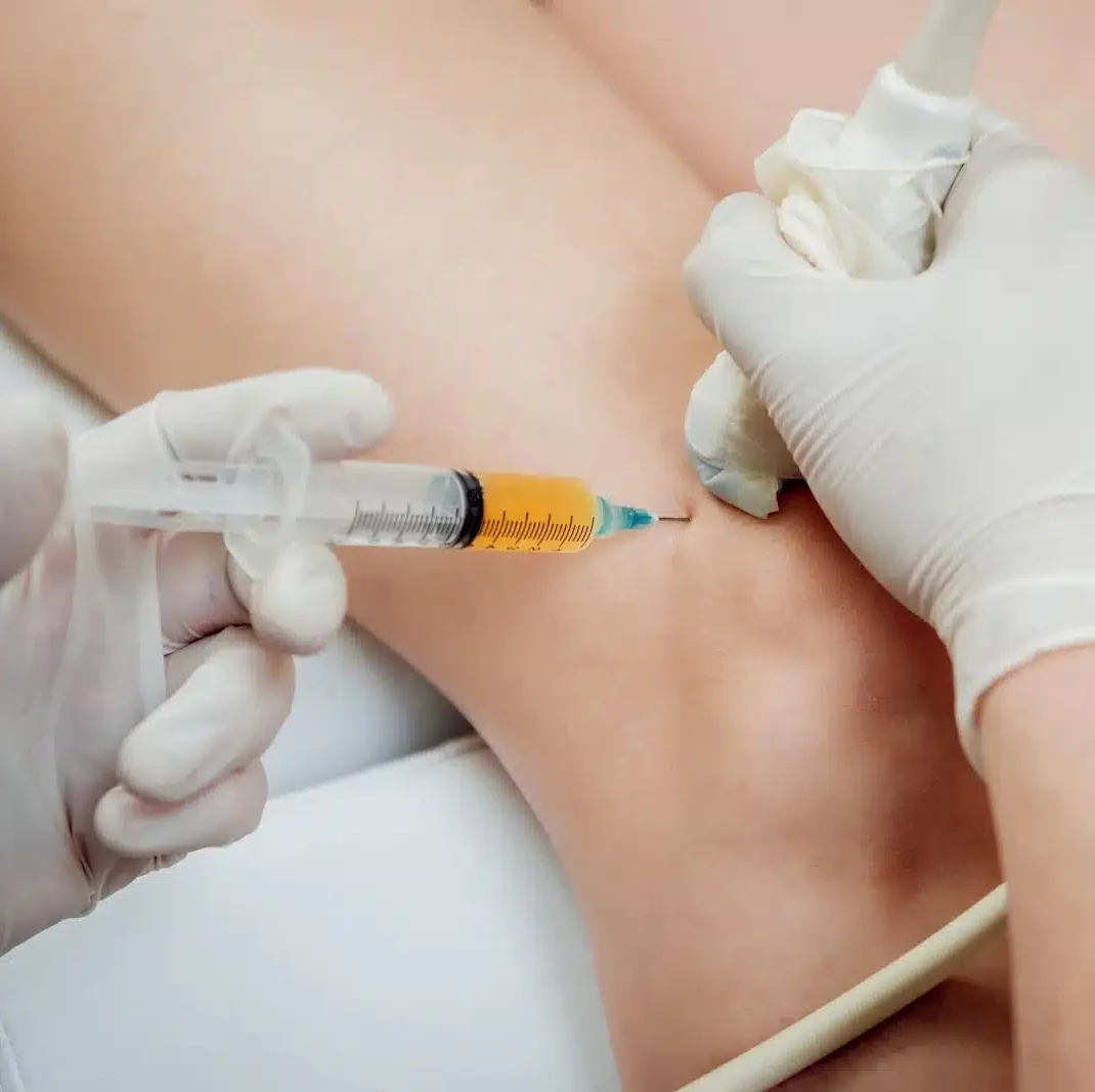
Yes. If the scan confirms a treatable problem, an ultrasound-guided hip injection can often be arranged: a trochanteric bursa injection for greater trochanteric pain syndrome, an iliopsoas bursa injection for iliopsoas bursitis, a hip joint injection for synovitis, a high-volume or PRP injection around a gluteal tendon, or aspiration of a bursa or collection.

Placing the needle under live ultrasound means it goes exactly where it is needed, which is more reliable than an unguided injection at the hip because the bursae and tendon insertions sit deep beneath thick soft tissue. Our one-stop orthopaedic and MSK clinic can take you from scan to treatment quickly. Treatment is booked and charged separately from the scan.

Where can I get a hip ultrasound near me in London?

Our Central London clinic is at 27 Welbeck Street, W1G 8EN, in the Harley Street Medical District, two minutes from Bond Street and a short walk from Oxford Circus, Marylebone and Baker Street, which covers most searches for a hip scan near me or a private hip ultrasound near me in central London. Because Bond Street is on the Elizabeth line, it is also a direct journey from Slough, Reading and the Thames Valley, and from Stratford and the east.

Our second clinic is at 54–56 Victoria Street, St Albans AL1 3HZ, a short walk from St Albans City station, serving Hertfordshire and North London. Both clinics run the same hip and upper thigh scan, £199 for one hip and £299 for both, seven days a week between 9am and 7pm.

Scroll to Top