Private Hernia Ultrasound Scan

Private Hernia
Ultrasound Scan

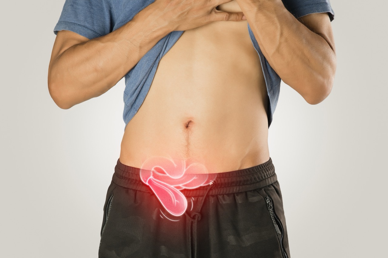
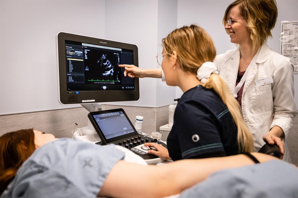
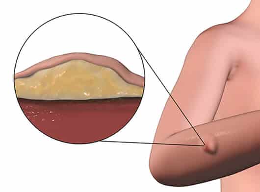
A rapid, painless and non-invasive ultrasound assessment for a suspected groin, inguinal, femoral, umbilical, incisional or abdominal-wall hernia. Dynamic ultrasound helps assess the area while resting, coughing or straining and can document the hernia’s location, size and behaviour.

No GP referral required
Same-day appointments often available
Written report after the scan or within 24 hours
Private Scan Price
£219
Targeted dynamic hernia ultrasound
Examines Groin, inguinal canal or abdominal wall
Duration Usually 15–20 minutes
Results Immediate discussion plus written report
Preparation No essential preparation
Book Online →
Assessment
Dynamic hernia scan
Areas
Groin or abdominal wall
Duration
Usually 15–20 minutes
Price
£219
Access
Self-referral available
What the Scan Includes

A focused ultrasound examination of the symptomatic or bulging area, performed dynamically to assess for a reducible or non-reducible hernia.

  • Review of your symptoms and relevant medical history
  • Ultrasound assessment of the suspected hernia site
  • Examination while resting and during cough or strain
  • Assessment of hernia location, size and contents where visible
  • Review of adjacent soft tissues and lymph nodes
  • Explanation of the findings during and after the examination
  • Official report with relevant images after the scan or within 24 hours
Common Reasons to Book

A scan can help investigate a lump, bulge, pressure or pain that becomes more noticeable with movement, coughing, lifting or standing.

  • A visible or palpable groin or abdominal-wall lump
  • Burning, aching or dragging discomfort
  • Pain when coughing, bending, lifting or straining
  • Weakness or pressure in the groin or abdominal wall
  • Swelling that changes between standing and lying down
  • Suspected recurrence after previous hernia surgery
  • An unclear lump that may have another soft-tissue cause
Private Scan Price
Hernia ultrasound scan: £219

Common hernia locations

The examination is tailored to the site of your symptoms or lump.

Inguinal Hernia

A groin hernia occurring through the inguinal canal. This is the most common hernia type.

Femoral Hernia

A hernia in the upper inner thigh or groin region, below the inguinal ligament.

Umbilical Hernia

A hernia at or close to the belly button.

Epigastric Hernia

A midline upper-abdominal-wall hernia between the breastbone and umbilicus.

Incisional Hernia

A hernia developing through or close to a previous surgical scar.

Spigelian Hernia

A less common lateral abdominal-wall hernia that may be difficult to identify clinically.

A targeted and dynamic assessment

Groin

Assessment of a painful, swollen or bulging groin region on one or both sides.

Inguinal Canal

Dynamic evaluation of the inguinal canal during rest, cough and strain.

Anterior Abdominal Wall

Assessment of a midline or lateral abdominal-wall lump or area of discomfort.

Surgical Scar

Review of a previous operative site where an incisional hernia is suspected.

Adjacent Soft Tissues

Assessment for alternative causes of a lump, including lymph nodes or soft-tissue lesions.

Clinical Next Steps

The report can support GP, surgical or specialist review and further imaging where appropriate.

How to prepare

There is no essential preparation for a standard hernia ultrasound scan.

1

Wear comfortable clothing

Choose clothing that allows easy access to the symptomatic groin or abdominal-wall area.

2

Identify the exact area

Be ready to point out the lump, pain or swelling and explain when it is most noticeable.

3

Bring previous information

Bring relevant surgical details, previous scan reports or referral letters when available.

Seek urgent medical care for possible strangulation

A suddenly painful lump that cannot be pushed back, especially with vomiting, abdominal swelling, fever, skin colour change or inability to pass stool or wind, may require emergency assessment rather than a routine private ultrasound appointment.

From booking to clear next steps

A straightforward private pathway with no GP referral required.

1
Book Online

Choose your preferred location and appointment time using the secure booking calendar.

2
Describe Your Symptoms

Explain the location of the lump or discomfort and when it becomes most noticeable.

3
Dynamic Ultrasound

The area is scanned while resting and, where appropriate, during coughing, straining or standing.

4
Receive Your Findings

The findings are discussed and documented in a formal report with relevant images.

5
Arrange Follow-Up

Where needed, the report can support GP review, surgical consultation or further imaging.

Frequently asked questions

The scan assesses the symptomatic groin, inguinal canal or abdominal wall for a hernia. It can document the site, size, contents and movement of the hernia and can also assess nearby soft tissues for another cause of the lump.
No essential preparation or fasting is normally required. Wear comfortable clothing and bring details of previous surgery or imaging when relevant.
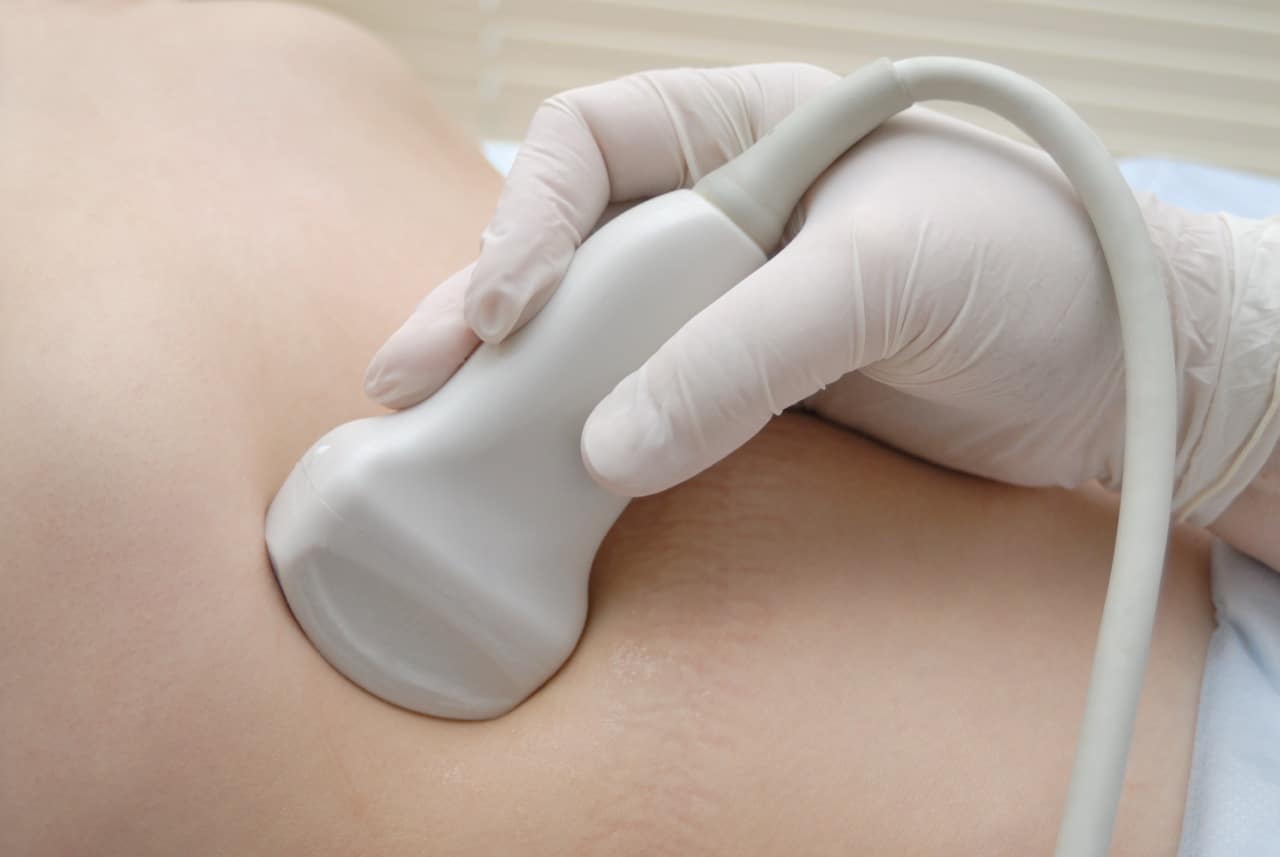
Water-based gel is applied to the area and the ultrasound probe is moved gently over the skin. You may be asked to cough, strain, change position or stand so the area can be assessed dynamically.
A standard hernia ultrasound is usually completed within approximately 15–20 minutes, although complex or multiple areas may take longer.
The findings are discussed verbally after the examination. The formal written report is prepared after the scan or within 24 hours and sent to you with any recommended actions.
Yes. Diagnostic ultrasound does not use ionising radiation and has no known routine side effects when used appropriately.

Book your hernia ultrasound scan

Select a location and convenient appointment time below.

Scroll to Top