
Can You See Cancer in the Abdomen With Ultrasound?
Ultrasound can show abnormalities in the abdomen that may turn out to be cancer, but it cannot confirm cancer on its own. This guide explains what abdominal ultrasound sees well, where it has limitations, and when CT, MRI or endoscopy may be more appropriate.
There are different types of imaging that can detect specific anomalies in the body.
Most people searching this question are somewhere between a symptom and an answer. There is pain that has not settled, weight that has come off without trying, an abnormal liver blood test, or a GP who has said the word scan without saying much else.
Regardless, we’re here to tell you that ultrasound can show abnormalities in the abdomen that may turn out to be cancer, including masses in the liver, kidneys, ovaries and lymph nodes.
But it cannot confirm a cancer diagnosis on its own.
So we researched this guide to let you know all about what an abdominal ultrasound sees well, and what it cannot see at all- basically all you need to know to answer the question you were googling.
What an abdominal ultrasound scan actually covers
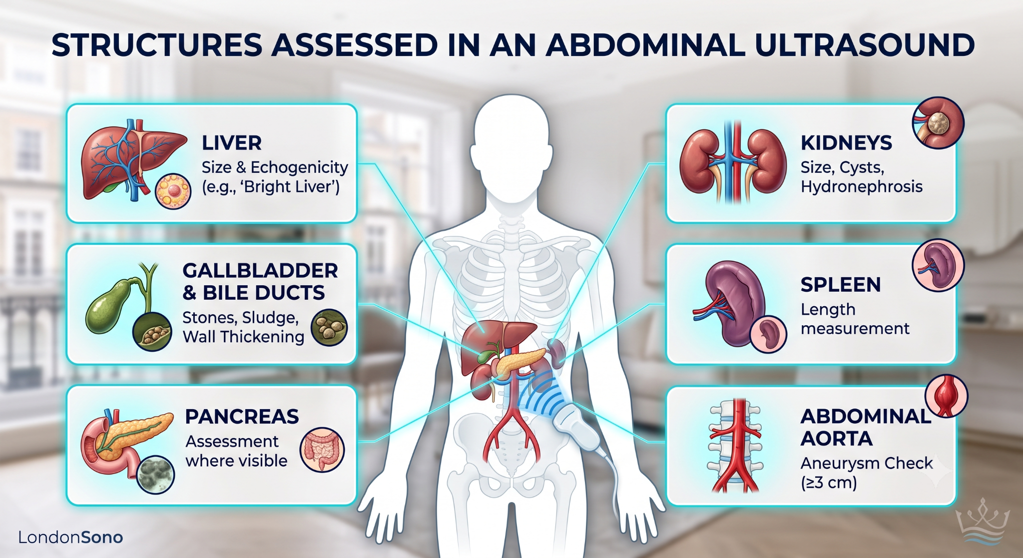
An abdominal ultrasound is a real-time scan of the upper abdomen using high-frequency sound waves. At London Private Ultrasound, we use it to assess the liver and liver texture, the gallbladder and bile ducts, the pancreas, both kidneys, the spleen and the abdominal aorta.
There is no radiation and no injection. The NHS states plainly that ultrasound scans do not use any radiation, unlike X-rays or CT scans.
Instead, the scan is looking at structure. It shows the size, shape, outline and internal texture of an organ, along with fluid collections, stones and solid lumps. Where a lump is found, ultrasound can often describe whether it is fluid-filled or solid, how well defined its edges are, and whether blood is flowing through it.
Those features are what a reporting clinician uses to decide whether something is recognisably harmless or needs a closer look.
What ultrasound can show, organ by organ
To really understand the type of information that an ultrasound can give us, here’s a quick breakdown of what we can actually see- by organs:
Liver
The liver is the abdominal organ that ultrasound visualises best. Because it’s large and sits directly under the ribcage, sound waves travel through it easily, allowing sonographers to detect a wide range of conditions with confidence.
Ultrasound can identify:
- Simple cysts (harmless fluid-filled sacs)
- Haemangiomas (benign clusters of blood vessels)
- Fatty liver changes
- The textured appearance of cirrhosis
- Focal lesions that look different from surrounding healthy tissue
- Secondary cancer deposits from elsewhere in the body, which often appear as multiple spots of varying brightness
When cancer from elsewhere in the body spreads to the liver, these patterns are recognisable to an experienced sonographer immediately. It’s worth noting that a single lesion in an otherwise normal liver is far more often benign than malignant, with simple cysts and haemangiomas being common incidental findings.
Gallbladder and bile ducts
Ultrasound is the first imaging test for gallbladder problems in almost every UK pathway. It shows gallstones clearly, along with wall thickening, polyps and a dilated common bile duct.
Gallbladder cancer is uncommon and usually presents as an irregular thickened wall or a mass replacing the gallbladder rather than as a discrete lump.
NICE recommends an urgent, direct access ultrasound scan to assess for gallbladder cancer in people with an upper abdominal mass consistent with an enlarged gallbladder.
Kidneys
Kidneys are well visualised on ultrasound, which can confidently distinguish between simple cysts and solid renal masses. This distinction matters because it guides whether further investigation is needed.
Ultrasound detects:
- Obstruction (blockages in urine flow)
- Kidney stones
- Scarring from previous infections or damage
- Solid masses that may require further investigation
Renal cell carcinoma is often found incidentally during scans requested for completely different reasons, which is one reason why kidney cancers are increasingly being detected while still small and more treatable.
Pancreas
The pancreas represents the most challenging part of an abdominal ultrasound examination, and it’s important to be straightforward about this limitation. The pancreas lies deep in the abdomen behind the stomach and transverse colon, and gas in either of these structures will block the sound beam.
While the head of the pancreas is usually visible, the body and tail often cannot be seen clearly. This is why, when pancreatic cancer is specifically suspected, CT scanning is typically the preferred test, with ultrasound serving as a starting point rather than a definitive answer. Our guide to what a pancreas ultrasound can assess goes through the anatomy and the conditions it picks up in more detail.
Spleen
The spleen is straightforward to image, and ultrasound can measure it accurately while showing various abnormalities.
The test can show:
- Accurate size measurements
- Cysts
- Infarcts (areas of tissue damage from blocked blood supply)
- Focal lesions
An enlarged spleen on its own doesn’t indicate cancer, but it’s a finding that prompts blood tests and sometimes further imaging since conditions like lymphoma and various blood disorders can cause splenic enlargement.
Abdominal aorta
One of the most consequential findings an abdominal ultrasound can detect has nothing to do with cancer at all. Ultrasound is the screening test for abdominal aortic aneurysm, and the NHS invites all men in England for a scan in the year they turn 65.
The test measures the aorta’s diameter and categorises any aneurysm as:
- Small: 3.0–4.4 cm
- Medium: 4.5–5.4 cm
- Large: 5.5 cm or bigger
This enables monitoring and intervention before a potentially life-threatening rupture occurs, making it one of the most important findings a routine abdominal scan can identify.
Lymph nodes
Enlarged lymph nodes around the aorta and at the porta hepatis (where blood vessels enter the liver) can be seen on ultrasound when they’re large enough, and bowel gas doesn’t block the view. The scan assesses the size, shape, and internal structure of visible nodes, with rounded nodes that have lost their normal fatty centre being treated as more suspicious.
It’s important to understand that lymph node assessment on ultrasound is inherently incomplete, since many abdominal lymph nodes sit behind bowel and cannot be visualised. This means a normal ultrasound doesn’t completely rule out lymph node involvement, and further imaging may be needed if there’s strong clinical suspicion.
Ovaries and pelvic organs
For women concerned about ovarian health, it’s important to understand that an upper abdominal ultrasound doesn’t assess the ovaries. A dedicated pelvic ultrasound is needed for this, and it’s the test recommended when ovarian cancer is suspected based on symptoms and CA125 blood test results.
Current guidance uses age-specific thresholds for the CA125 blood test to determine when an urgent ultrasound should be arranged, with different values for different age groups. This approach helps catch ovarian cancer earlier while accounting for the fact that CA125 can be elevated for many non-cancerous reasons.
What an abdominal ultrasound cannot show
Understanding what an abdominal ultrasound cannot show is as important as knowing what it can. Sound does not travel through gas or bone, and it loses resolution with depth. Everything on this list follows from those two facts.
- The stomach and bowel lining. Gas blocks the beam, so the surfaces where stomach and bowel cancers begin are not reliably assessable. Endoscopy and colonoscopy are the tests that look at them.
- Much of the pancreas in many people. The body and tail are frequently obscured by overlying gas, and a small tumour can be missed entirely.
- Small liver lesions. This is where the published numbers are uncomfortable. A 2018 meta-analysis in Gastroenterology by Tzartzeva and colleagues found that ultrasound surveillance detected hepatocellular carcinoma at any stage with 84% sensitivity, and early-stage disease with only 47% sensitivity.
- Anything sitting behind bone or lung. The upper dome of the liver lies under the ribs and diaphragm, and part of it can be hidden from view.
- Fine detail at depth. Sound attenuates as it travels, so image quality falls as the distance from the probe to the organ increases.
- Whether something is cancer. Ultrasound describes appearance. Cancer Research UK notes that one of its roles is to guide doctors when they need to take a tissue sample, and tissue is what confirms a diagnosis.
A normal abdominal ultrasound is reassuring, and it is not the same as a clear result for the whole abdomen. It means nothing abnormal was seen in the organs the scan can assess, on the day of the scan, at the image quality available. If your symptoms continue or change, tell a clinician, whatever the report said.
Ultrasound compared with CT, MRI and endoscopy
| Test | How it works | Radiation | Best for | Less suitable for |
|---|---|---|---|---|
| Ultrasound | Sound waves, real-time imaging | None | Liver, gallbladder, kidneys, spleen, aorta, ovaries, testes, superficial lymph nodes | Pancreas body and tail, stomach, bowel, lungs, anything behind gas or bone |
| CT | X-rays from many angles, reconstructed | Yes | Staging, pancreas, bowel wall, lungs, a whole-abdomen overview in one pass | Repeated use in younger patients, characterising some liver lesions |
| MRI | Magnetic fields and radio waves | None | Characterising liver lesions, bile ducts (MRCP), pelvic soft tissue | Cost and availability, some implants, claustrophobia |
| Endoscopy and colonoscopy | A camera passed into the gut | None | The lining of the stomach and bowel, with biopsy in the same appointment | Anything outside the gut lumen |
Swipe horizontally to view the full table.
Our fuller comparison of abdomen ultrasound and other imaging techniques works through the same tests one by one.
London Private Ultrasound provides ultrasound, Doppler, echocardiography, ECG, blood tests and consultations. Based on your result and if you need a follow-up or any other test, such as an MRI- we refer you to a GP at London Private Ultrasound- at an additional cost.
When NICE says an ultrasound is the right next step
In the UK, GPs follow NICE guideline NG12 (Suspected cancer: recognition and referral), which names ultrasound directly in several places. The guideline was most recently updated in April 2026.
Where ultrasound is the first-choice test
Ultrasound is the named test for several specific concerns where it’s the appropriate first investigation.
Gallbladder and liver: An upper abdominal mass consistent with an enlarged gallbladder or liver should be assessed with an urgent direct-access ultrasound.
Ovarian cancer: For women aged 40 and over with persistent symptoms, GPs measure CA125 and arrange urgent ultrasound if results reach age-specific thresholds (ranging from 35 IU/mL for ages 40–49 down to 24 IU/mL for ages 60–69). For women under 40, ultrasound is considered directly without relying on CA125.
Endometrial cancer: Women aged 55 and over with unexplained vaginal discharge should be considered for direct-access ultrasound.
Testicular cancer and soft tissue sarcoma: Urgent ultrasound is recommended when these are suspected.
Where ultrasound has limitations
Pancreatic cancer: CT scan is the first choice. Ultrasound is only recommended if CT is not available, reflecting the pancreas’s difficult position deep behind the stomach and bowel.
What this means if you’re booking privately
Ultrasound is the first-line test for gallbladder, liver, ovarian, endometrial and testicular concerns. If you’re booking privately because an NHS wait is long, this tells you which concerns a private ultrasound genuinely addresses. For pancreatic concerns specifically, CT would be more appropriate.
What happens if the scan finds something
This is the question behind the question, and it’s worth answering directly.
Most findings fall into one of three categories. The largest by far is recognisably benign: simple cysts, haemangiomas, gallstones, fatty liver. These are described in your report and usually need reassurance or a routine repeat scan, nothing more.
Some findings are indeterminate — the scan has seen something real but can’t characterise it with confidence. The next step is usually a different test (often CT or MRI) or a repeat ultrasound after a few months to see whether anything has changed.
A smaller group of findings have features that raise concern. Here the pathway matters more than the scan itself. In England, the NHS faster diagnosis standard expects people referred with suspected cancer to have a diagnosis confirmed or ruled out within 28 days of referral.
Where a private scan finds something that needs NHS specialist management, we produce a referral letter the same day at no additional charge because it carries confirmed imaging findings, that referral usually gives your specialist more to work with at the first appointment.
How to read your abdominal ultrasound report
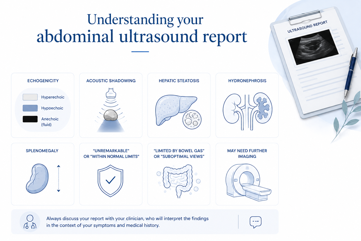
Here’s what the usual terminology on your report might mean:
| Term in your report | What it usually means |
|---|---|
| Lesion | Any area that looks different from the tissue around it. This is a neutral word with no implication of cancer — it simply means “something we noticed that looks distinct”. |
| Mass | A solid area taking up space. It needs characterising, and many turn out to be benign. The word sounds worrying, but it’s descriptive rather than diagnostic. |
| Cyst | A fluid-filled sac. Simple cysts are very common in the liver and kidneys and are almost always harmless. They’re one of the most frequent incidental findings on abdominal scans. |
| Hypoechoic or echogenic | Darker or brighter than the surrounding tissue on the image. This is purely a description of how something appears on the scan, not a diagnosis of what it is. |
| Indeterminate | Something was seen, but it couldn’t be confidently characterised on this scan. Another test (often CT or MRI) normally follows to get a clearer picture. |
| Incidental finding | Something the scan found that it wasn’t originally looking for. These are extremely common and often turn out to be nothing serious. |
| Clinical correlation advised | This finding should be read alongside your symptoms and blood results before deciding what to do next. It means the scan alone doesn’t tell the whole story. |
Swipe horizontally to view the full table.
How to prepare for an abdominal ultrasound
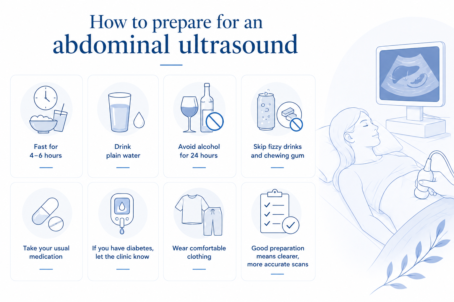
Here’s a quick overview of how to prepare for an abdominal ultrasound:
- Fast for 4 to 6 hours before your appointment.
- Avoid food, tea and coffee during the fasting period. Small sips of water are fine.
- Avoid fatty food and fatty drinks, which empty the gallbladder and make it harder to assess.
- Avoid fizzy drinks and chewing gum, both of which increase gas in the stomach.
- Wear loose clothing that gives easy access to your abdomen.
- Bring any previous scan reports or recent blood results.
How much does a private abdominal ultrasound cost in London?
At London Private Ultrasound an abdominal ultrasound scan is £219. That covers assessment of the liver and liver texture, gallbladder and bile ducts, pancreas, kidneys, spleen and abdominal aorta, a consultation with your sonographer, an immediate verbal explanation of the findings, and a written report usually within 24 hours. The Private Abdominal and Pelvic Ultrasound Scan is £399, and the Liver Health Package is £449.
Across central London, private abdominal ultrasound prices run from roughly £150 to £300. Some clinics advertise a lower headline figure for a scan limited to a single organ, and some quote off-peak rates that only apply outside weekday daytime slots.
What to check before comparing prices
- Whether a written report is included in the price or charged as an extra.
- Who performs and reports the scan, and their professional registration.
- How long the written report takes to arrive.
- How many organs the advertised price actually covers.
- Whether a referral letter is included if something needs following up.
Book a private abdominal ultrasound at London Private Ultrasound
Let’s be honest — most people book with us because they’re tired of waiting. Maybe your GP suggested a scan weeks ago, and you’re still on the list. Maybe you’ve got symptoms that are making you anxious, and you just want answers. Whatever the reason, we get it. The waiting is often worse than the scan itself.
Here’s a couple of reasons why our patients choose us:
Same-day appointments: We know that when you’re worried, you don’t want to wait. We offer same-day and next-day slots seven days a week, 9am to 7pm.
No GP referral needed: Book directly without waiting for your GP to arrange anything. Call, WhatsApp, or book online.
Expert sonographers: Every scan is done by a registered ultrasound consultant or sonographer with years of experience.
Results on the day: You’ll get verbal results before you leave, plus a written report within 24 hours.
A welcoming team: Scans can feel daunting, especially if you’re worried. Our team takes time to explain what’s happening, answer your questions, and make sure you feel comfortable throughout.
If your report mentions that you need a GP referral or further investigation, we’ll provide that referral letter for you. We can also book a follow-up consultation with one of our doctors if needed (at an additional cost), so you’re not left figuring out next steps on your own.
Frequently Asked Questions
Can an ultrasound scan detect cancer?
An ultrasound scan can show abnormalities that may turn out to be cancer, such as a solid mass in the liver or a solid renal lesion. It cannot confirm a cancer diagnosis on its own. Confirmation comes from a tissue sample, and Cancer Research UK describes one of ultrasound’s roles as guiding doctors to the right place to take that sample.
Can an abdominal ultrasound miss cancer?
Yes. Ultrasound cannot see through gas or bone, so the stomach lining, the bowel and much of the pancreas are poorly assessed. A 2018 meta-analysis in Gastroenterology found ultrasound detected early-stage liver cancer with 47% sensitivity. A normal scan lowers the likelihood of some cancers and does not exclude all of them.
Does a normal abdominal ultrasound mean I do not have cancer?
No. It means nothing abnormal was seen in the organs the scan assessed, on that day, at the image quality available. If your symptoms persist or change, speak to a clinician regardless of the result.
What does it mean if my report says lesion?
Lesion is a neutral radiology word for an area that looks different from the tissue around it. Most lesions found on an abdominal ultrasound are benign, including simple cysts and haemangiomas in the liver. Your report will say whether anything further is needed.
Can ultrasound see pancreatic cancer?
Sometimes, and it is unreliable for it. The pancreas lies deep in the abdomen behind the stomach, and the body and tail are frequently obscured by gas. NICE recommends urgent, direct access CT for suspected pancreatic cancer, with ultrasound as the alternative when CT is not available.
Can ultrasound see bowel or stomach cancer?
Not reliably. Gas in the stomach and bowel blocks the sound beam, so the linings where these cancers begin are not assessable. Endoscopy and colonoscopy are the tests that look at those surfaces directly, and they allow a biopsy at the same time.
Do I need a GP referral for a private abdominal ultrasound?
No. You can self-refer and book directly. Bring any previous scan reports or blood results if you have them, as they give your sonographer useful clinical context.
Is an abdominal ultrasound safe?
Yes. Ultrasound uses sound waves rather than ionising radiation. The NHS states that ultrasound scans do not use any radiation, unlike X-rays and CT scans, and the scan can be repeated as often as is clinically appropriate.
How long does an abdominal ultrasound take, and when do I get results?
The scan itself takes around 20 minutes, with extra time for your clinical history and an explanation afterwards. You receive a verbal explanation of the findings immediately, and a written report usually within 24 hours.
Do I need to fast before an abdominal ultrasound?
Yes. Fast for 4 to 6 hours beforehand, and avoid food, tea, coffee, fatty drinks, fizzy drinks and chewing gum. Small sips of water are fine. Fasting keeps the gallbladder full and reduces gas, which is what makes the gallbladder and pancreas assessable.
How is a private abdominal ultrasound different from an NHS one?
The technique and the equipment are the same. What differs is timing and the report route. Privately you choose the appointment, often on the same day, and the written report comes back to you directly. Booking privately does not affect your NHS care, and a referral letter is provided if NHS specialist management is needed.
What happens if the scan finds something?
Findings fall into three groups: recognisably benign, indeterminate, or concerning. Benign findings usually need reassurance or a routine repeat. Indeterminate and concerning findings lead to a further test or a specialist referral, and we issue a referral letter the same day at no extra charge.
Clinical sources:
Need clear answers about abdominal symptoms?
A private abdominal ultrasound scan is £219, with consultation, verbal findings on the day and a written report usually within 24 hours. No GP referral is required.