
Varicocele: Symptoms & How It Is Diagnosed on Ultrasound
A varicocele is a group of enlarged veins around a testicle. Ultrasound can measure the veins, assess reversed blood flow and compare both testicles to help guide the next step.
Varicocele is more common than many people realise. Research suggests that around 15–20% of adult men have one—roughly three men in a room of twenty—but most will never know unless symptoms or a fertility investigation prompt a scan.
Patients usually come to us when discomfort warrants assessment or when difficulty conceiving makes a men’s fertility check-up appropriate. This guide explains varicocele, when to consult a GP and how ultrasound helps with diagnosis.
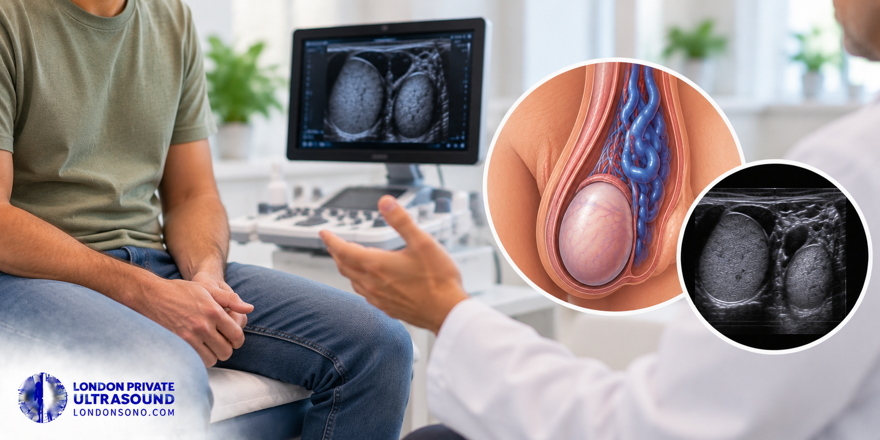
A large varicocele may feel like a “bag of worms” above the testicle. Smaller varicoceles may not be felt and are found only on a scan.
Why are varicoceles almost always on the left?
Varicoceles occur on the left in 80–90% of cases because of how the veins drain on that side. The left testicular vein joins the left renal vein at a right angle, creating more back pressure than on the right, where the vein drains at a gentler angle into the main abdominal vein.
If you have a left-sided varicocele, there is a 30–40% chance that a scan will also find one on the right. An isolated right-sided varicocele is less common and may require assessment of the abdomen because pressure on the vein can sometimes be responsible.
Where appropriate, we may recommend a kidney and urinary tract ultrasound to help investigate the underlying cause.
What are the symptoms of a varicocele?
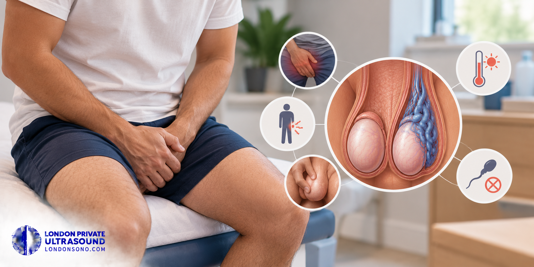
Most varicoceles cause no symptoms. When symptoms do occur, they may include:
- A dull, dragging ache in the scrotum, usually on one side.
- Discomfort that builds during the day and is worse after standing, exercise or hot weather.
- A visible or palpable lump above the testicle that softens when you lie flat.
- A testicle that looks or feels smaller on the affected side.
- Difficulty conceiving, which may be the only reason a varicocele is found.
How is a varicocele diagnosed?
A varicocele is assessed by clinical examination and confirmed with a scrotal ultrasound. A clinician examines you standing, both at rest and while you bear down—known as the Valsalva manoeuvre.
Swipe sideways to view the full table.
| Grade | What the examination finds |
|---|---|
| Grade 1 | Felt only while bearing down. |
| Grade 2 | Felt without bearing down, but not visible. |
| Grade 3 | Visible through the skin of the scrotum. |
| Subclinical | Nothing felt on examination; seen only on ultrasound. |
Book a private testicular ultrasound
A scrotal ultrasound at our Central London or St Albans clinic measures the veins, uses colour Doppler during Valsalva and compares both testicles. The scan usually takes 10–15 minutes, and the clinician explains the findings. No GP referral is required.
Book a Testicular UltrasoundWhat does an ultrasound show in a varicocele?
A testicular and scrotal ultrasound scan measures the veins and shows the direction of blood flow. It assesses:
- Vein diameter: European ultrasound guidance uses 3 mm or more as a diagnostic threshold when measured standing and during Valsalva.
- Reflux: This means blood flowing backwards. A threshold of more than two seconds of reversed flow is used, measured standing and during Valsalva.
- Testicular volume: Both sides are measured. A difference greater than 20% matters particularly in teenagers and is one accepted reason to consider treatment.
- Both sides: Bilateral varicoceles are common and may change the treatment plan.
- Other scrotal findings: The scan also checks for hydroceles, epididymal cysts and solid lumps.
What else could be causing scrotal swelling or ache?
Several conditions can cause scrotal swelling or pain. Ultrasound is usually the best first diagnostic test to distinguish between them.
Swipe sideways to view the full table.
| Condition | How it differs |
|---|---|
| Hydrocele | A smooth fluid collection around the testicle that transmits light and does not change with position. |
| Epididymal cyst or spermatocele | A discrete, round lump behind or above the testicle, usually painless and separate from it. |
| Epididymitis | Develops over days with tenderness, redness and sometimes fever or urinary symptoms. |
| Inguinal hernia | A swelling extending from the groin into the scrotum, often more obvious with coughing or standing. |
| Solid testicular lump | Any firm lump arising from the testicle itself requires prompt assessment. |
If a firm lump is found within the testicle, prompt assessment is important. Our Testes Health Check combines ultrasound with relevant blood tests where appropriate.
Does a varicocele affect fertility?
A varicocele can affect fertility and is the most common potentially correctable finding in men being investigated for infertility. It may increase the temperature around the testicle and affect sperm production, so sperm count, movement and shape can be reduced.
For a couple whose investigations identify a varicocele, treatment can be a reasonable option with a chance of improving fertility. The decision should be made with a fertility specialist who can consider the semen analysis, the partner’s assessment, symptoms and scan findings together.
What are the treatment options for a varicocele?
Treatment usually falls into three groups:
Suitable when there are no symptoms, semen parameters are normal and there is no size difference between the testicles. Supportive underwear and simple pain relief may help mild aching.
An interventional radiologist passes a fine catheter through a vein and blocks the affected vein with coils so blood is redirected. It avoids general anaesthesia and recovery is usually quick.
Microsurgical varicocelectomy ties off the affected veins through a small groin incision, usually under general anaesthesia.
Treatment is generally considered for a palpable varicocele with abnormal semen parameters, persistent pain, problematic bilateral varicoceles, or an adolescent with more than a 20% difference in testicular size.
London Private Ultrasound can diagnose, grade and measure a varicocele, then refer you to a private GP appointment, urologist, interventional radiologist or fertility specialist according to the scan and symptoms.
Can a varicocele be cured completely?
In most cases, a varicocele can be treated successfully and most men who have a procedure obtain lasting symptom relief. Recurrence occurs in up to 10% of cases because small collateral veins can open and take over from the treated veins.
Book a testicular ultrasound at London Private Ultrasound
If you have a scrotal ache, a lump above a testicle or an ongoing fertility investigation, a scrotal ultrasound provides measurements that can help determine the next step.
London Private Ultrasound has been scanning patients since 2012 and is CQC registered. Our clinics are in Central London and St Albans, with appointments available seven days a week from 9am to 7pm.
No GP referral is needed. Call 020 7101 3377 or book online.
Arrange your assessment
Book a private testicular ultrasound for an expert assessment, colour Doppler measurements and a written report.
Frequently asked questions
How much does varicocele surgery cost in the UK?
How long is the NHS wait for varicocele treatment?
Does ejaculating reduce a varicocele?
Can a varicocele go away on its own?
Should a teenager with a varicocele be treated?
Can I lift weights or exercise with a varicocele?
Does a testicular ultrasound hurt, and do I need a referral?
Does a varicocele cause low testosterone?
Clinical disclaimer: This article provides general health information and does not replace individual medical advice, examination or diagnosis. Seek urgent medical attention for sudden severe testicular pain, rapid swelling, fever or a firm testicular lump.
Medical review: Mr Pedram Aghaei, Co-founder and Medical Writer, London Private Ultrasound. Content reviewed against current NHS, NICE and professional ultrasound guidance. Review cycle: every six months.