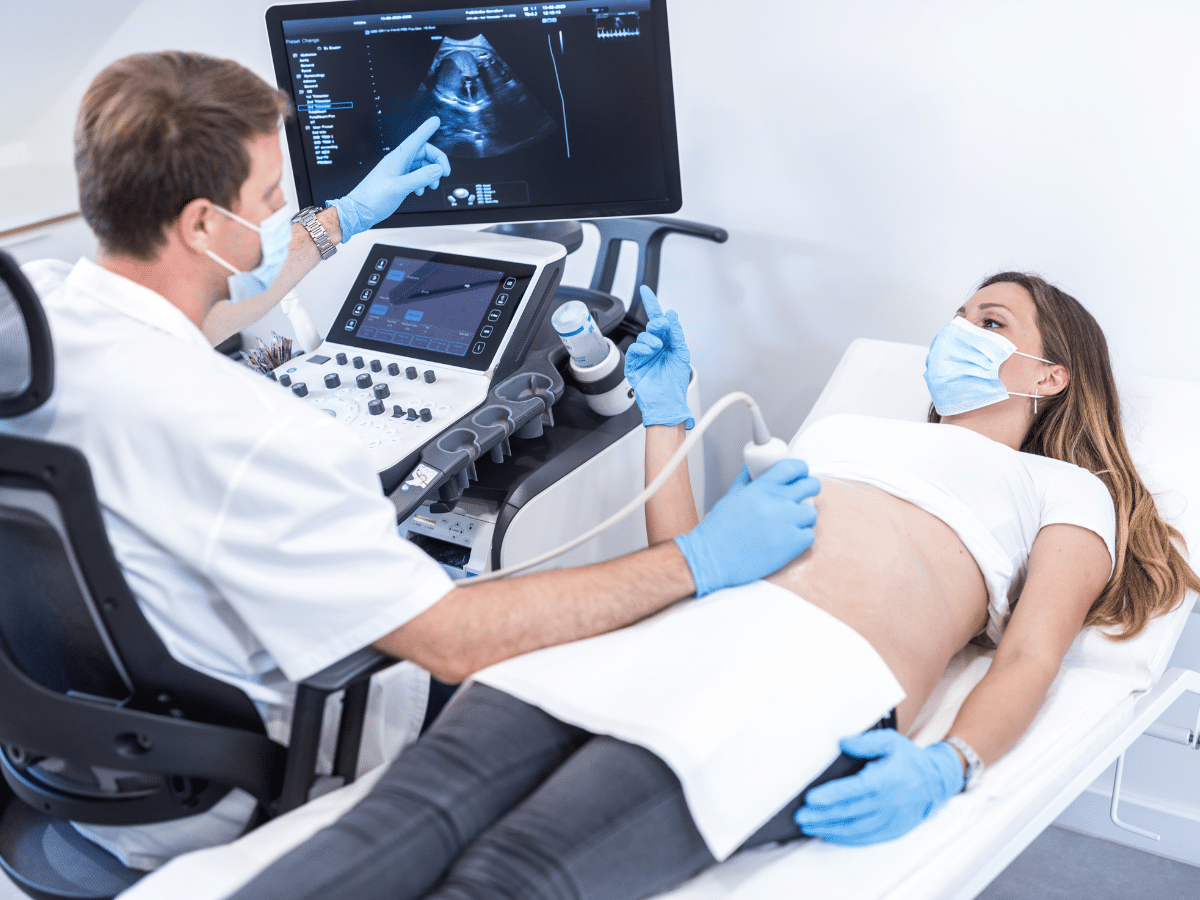
Ultrasound's Role in diagnosing First-trimester Pregnancy Issues
Ultrasound is a sensitive tool for the diagnosis of fetal anomalies during the prenatal period. Routine ultrasound examination has been a recognized element of antenatal care. Being increasingly offered during the first semester, however it is commonly performed in the second trimester. (Bau and Atri, 2000; Papp and Fekete, 2003). In fact, it has become the main imaging tool and the first line of diagnostic investigation in first trimester pregnancy examinations. Supported by the fact that it is non-invasive (Okaro and Valentin, 2004) and produces real time, immediate results, it helps to triage patients into appropriate management protocols. Recent massive technological developments such as high-frequency transvaginal scanning, have made huge progress in the resolution of ultrasound imaging in the first trimester with which early fetal development can be comprehensively evaluated and monitored.
NORMAL PELVIC ANATOMY
The normal female reproductive system comprises of vagina, uterus, fallopian tubes and ovaries and on ultrasound their appearance is dependent on patients’ age and hormonal status at the time of imaging (Bates, 2006). The vagina, which is a thin walled muscular structure lies midline and appears as a hyperechoic midline echo. From the vagina, the uterus, a pear shaped muscular organ can be followed. This also lies in the midline and can be anteverted, retroverted or retroflexed (Bates, 2006b, Levi et al, 2008). The uterine wall is composed of three layers, each exhibiting its own characteristics on ultrasound (Alty and Hoey, 2006). The outer parametrium is very echogenic and outlines the uterus, the intermediate layer, the myometrium is less reflective and gives homogenous echoes while the endometrium which is the innermost layer changes its thickness (pre-menopause <15mm, post menopause <5mm) and reflectivity due to hormonal status of the patient. The ovaries are small oval shaped organs lying posterolateral on either side of uterus. Ovarian follicles are seen as a well-defined echo free areas that change in size with the menstrual cycle, varying from 2mm-20mm in diameter (Levi et al, 2008). The fallopian tubes, peritoneal recesses and ligaments are not visualized on ultrasound unless surrounded by, or contain fluid (Levi et al,2008).
TECHNIQUE
The standard first trimester pregnancy ultrasound examination involves transabdominal (TAS) and transvaginal (TVS) approaches, if there are no contraindications (Levi, et al 2008). A curvilinear 3.5-5MHz probe and a transvaginal high frequency probe usually a 7.0 to 8.0 MHz are required.
TAS must be done with a full urinary bladder, it gives a wider field of view providing better visualization and measurement of gestational sac toward the uterine fundus and other structures in the pelvic abdominally. To measure the maximum longitudinal axis of the sac sliding the probe and / or rotating slightly to either side is helpful (Levi et al, 2008).
As Bates points out, TAS is also useful in locating ovaries in relation to uterus, picking up uterine anomalies and scanning the iliac fossae and bladder. Levi et al. (2008) argue that this is limited by patients’ body habitus. TAS should be performed in a true longitudinal segment of the uterus, if it is not possible to visible the gestational sac, panning the transducer is essential from side to side until the whole of the uterus has been examined and the maximum length of the gestation sac is displayed.
A further disadvantage of TAS is the fact that some patients cannot tolerate the distended bladder or else because of coexisting morbidities cannot reach adequate bladder filling (Bates 2006a). TVS on the other hand allows detailed examination of pelvic organs without requiring the bladder to act as an acoustic window. Therefore, TVS should be performed in all situations when there are no contraindications (Levi et al, 2008).
As pointed out by Bates (2006a) TVS disadvantages include narrower field of view, decreased probe flexibility during scanning, acceptability by patients who may regard it as invasive and uncomfortable, however this is generally overcome by the sonographer’s good communication skill and ensuring patients’ privacy and dignity (Bates, 2006a).
ROLE OF FIRST-TRIMESTER ULTRASOUND
Performing the first-trimester ultrasound either as a routine ultrasound study between 10 – 13 weeks gestation or for patients presenting abdominal pain, vaginal bleeding or both are recommended by the International Society of Ultrasound in Obstetrics and Gynaecology (ISUOG) (Salomon et al. 2013). In assessing of the early pregnancy complications such as miscarriage, molar or ectopic pregnancy, ultrasound is gold standard diagnostic modality (Chandrasekaran and Thilaganathan, 2012). Although differentiating of the findings and accuracy of the ultrasound scan is depends on gestational age and operators experience.
A pregnancy is considered ongoing only if an embryo with cardiac activity and CRL≥7.0 mm is visible within the GS≥25.0mm according to the current ISUOG guidelines. Absent of embryonic heartbeat, empty/growth inconsistencies in GS size or CRL strongly advocate a nonviable pregnancy (Datta, 2013). A bradycardic embryo, underdeveloped GS or CRL, atypical yolk sac and sub-chorionic haematoma can be a primary sign of miscarriage (Bottomley and Bourne, 2009b). To differentiate a pregnancy of unknown viability from miscarriage and assists selecting the appropriate management protocols restricted criteria revised by RCOG (Bourne and Bottomley, 2012).
Additionally, the classification of miscarriage is based on the ultrasound findings (Jurkovic et al.2009). Colour Doppler also can facilitate differential diagnosis by showing perfusion (Chudleigh, 2016). Molar pregnancies have variable sonographic appearances and ultrasound can determine its type and extent (Kirk et al. 2007). Ectopic pregnancy is a challenging condition suspected when the endometrium is thickened or has irregular echogenicity, and no intrauterine sac (Chandrasekhar, 2008). Operators shouldn’t be confused to differentiate ectopic pregnancy with an avascular, irregular-shaped, pseudo-sac or a corpus luteum with circumferential blood flow (Hourani et al. 2008). The 3D ultrasound and 3D power Doppler improved detection of the site of EP and detailed vascularization analysis (Kirk, 2012).
Routine first-trimester ultrasound has been validated for accurate pregnancy dating, fetal number and chorionicity determination in multiple pregnancies, aneuploidy screening, detecting fetal structural abnormalities, and guiding fetal therapy (Chandrasekaran and Thilaganathan, 2012; Kurjak and Chervenak, 2011).
Ultrasound performed at 10-13+6 weeks’ gestation alongside confirming location and viability, determines the gestational age by measuring the CRL. The LMP is also useful only if it is known and regular, the UK National Institute for Clinical Excellence guideline on antenatal care advocate dating by CRL as the most precise method. For assessing the true gestational age care must be taken to measure the CRL correctly and when CRL measures more than 84mm, alternative sonographic measurements like femur length and abdominal or head circumference shown comparable accuracy (Bottomley and Bourne, 2009a).
To sonographically establish and detect multiple pregnancies, it is essential to look at the number of placentas and the inter-twin membrane thickness, and revealing the lambda or T-sign. Distinguishing chorionicity has a prognostic value for consequent development of discordant growth or/and twin-to-twin transfusion syndrome (Alhamdan et al. 2009). In addition, the necessary information regarding the vanishing twin phenomena, conjoined twins, and foetal abnormalities would be available to diagnose an early ultrasound assessment of twin pregnancy (Constantine and Wilkinson, 2015). Furthermore, as twinning is linked with high perinatal mortality, early ultrasound can provide an appropriate growth monitoring and if required, assists therapeutic interventions (Mukherjee and Thilaganathan, 2010).
Recent evidence suggests that ultrasound at 11-13weeks’ gestation for nuchal translucency (NT) measurement combined with maternal age, serum B-HCG and PAPP-A identifies foetuses affected by trisomy 13, 18 or 21, providing patient-specific aneuploidy risk assessment profiles (Norton, 2017). Structural defects (including major cardiac defects, diaphragmatic hernia) and genetic syndromes can be detected by noticing any increased in NT thickness (≥3.5mm) (Breeze et al. 2011). However, time of the measurement and diagnose is vital here as it can affect the result to be inaccurate and abnormalities to be missed. It is difficult to differentiate if a specific minor marker occurred by a normal variant or it is a sign of aneuploidy (Kurjak and Chervenak, 2011). NT refers to a collection of subcutaneous anechoic, fluid-filled space behind the foetus neck (Dulay and Copel, 2008) and conducting the test provides earlier prenatal diagnosis.
Congenital anomalies including defects of the central nervous system, heart, anterior abdominal wall and urinary tract are detectable in ultrasound between 11-14weeks′ gestation (Blaas, 2014; Roberts and Bhide, 2007). Some of the structural abnormalities can occur as isolated events and an increased NT indicates more detailed fetus scanning. There is an argument about the various structural abnormalities which can be reliably detected in first trimester and whether, a termination is appropriate or not (e.g. anencephaly, which is not compatible with life) (Moore and Bhide, 2009). Norton (2017) believes detection of structural and chromosomal abnormalities is essential in the first-trimester ultrasound, but it should never substitute for the second-trimester scan.
Amniocentesis and chorionic villus sampling (CVS) are two available invasive methods for obtaining a prenatal karyotype. Amniocentesis is normally performed after 16weeks, due a significantly higher risk of clubfoot, amniotic fluid leakage or foetal loss, when done earlier (Chisholm and Ferguson, 2012). The CVS sampling normaly performed for woman with a positive screening test, at 11-13weeks′ gestation. Under ultrasound guidance, the sampling path and localization of placenta can be determined (Chisholm and Ferguson, 2012; Coroyannakis and Thilaganathan, 2015).
ADVANTAGES AND LIMITATIONS
However, the pulsed and colour Doppler help more accurate fetal circulation assessment and improve growth survey (Kupesic and Kurjak, 2011), because of the potential risks, they should be avoided. Direct visualization of the heart in B mode or M-mode are the best ways to monitor heart activity and measure its rate (Wladimiroff and Eik-Nes, 2009).
3D/4D ultrasound adds clinical value to 2D ultrasound due to more accurate volume acquisition and increasing the rate of fetal anomaly detection (Sheiner et al. 2007; Vaughan, 2009). Developed ultrasound applications such as Doppler for assessing the uterine artery has increased the accuracy of routine sonographic examinations, however, there are still some safety issues (Ter Haar et al. 2013).
During the first trimester of pregnancy other cross imaging modalities like MRI are not suggested due to possible harmful effects on vulnerable embryo/foetus (Reddy et al. 2008). Jurkovic (2009) has suggested using MRI scanning occasionally for women with an unsure diagnosis of interstitial ectopic pregnancy, only if 3D scanning is not available.
Considering all above facts, sonography is still the gold standard for first trimester investigation. Although, there are still some weaknesses in ultrasound scanning such as operator competency, technical equipment and pitfalls like obesity with serious effects on foetuses’ anomaly detection rate.
SAFETY
Regardless of numerous privileges for ultrasounds safety, its helpfulness requires careful consideration of potential thermal or mechanical bio-effects (Abramowicz, 2013).
A TVS potentially can have more thermal and mechanical bio-effect due to tissue-probe closeness, using less intensity than TAS can control negative effect on foetus development and growth (Norton, 2017). Furthermore, on-screen safety indices (mechanical and thermal index) assist sonographer’s judgement. Consequently, international professional bodies approve that in the face of evidence of harm effect of Doppler examinations and raise in energy outputs, thereby safety cannot be acknowledged and it should only be use if clinically indicated (Salvesen et al. 2011; Ter Haar et al. 2013).
CONCLUSION
From the discussion, above, one can see that ultrasound is a valuable tool in first trimester pregnancy diagnosis and management with benefits outweighing risks if performed by well trained professionals. A combination of technical abilities, clinical knowledge and skills is required to reach accurate diagnosis.
A well prepared cooperative patient also contributes to successful outcome. Regular quality control procedures and auditing are a must for upkeep of good quality service. Considering the above evidence to provide good safe practice, a sonographer should;
– Acquire recognized training, keeping skills updated with continuous practice and CME, Use ultrasound as an extension of clinical practice not as replacement
– Perform each examination appropriately making an informed choice for patient preparation, procedure and equipment, Perform regular quality control procedures and audit.
– Use protocols and guidelines Respect the ALARA principle, as the basic safety principle to protect patients from over exposure.
Why Choose London Private Ultrasound?
At London Private Ultrasound, we prioritize your health and comfort. Our team of experienced radiologists and healthcare professionals are dedicated to providing the highest quality of care. Here’s why you should choose us:
- Expert Radiologists: Our team consists of highly trained and experienced radiologists who are experts in ultrasound diagnostics.
- State-of-the-Art Technology: We use the latest ultrasound machines to ensure the highest level of accuracy in our scans.
- Patient-Centered Care: We focus on creating a comfortable and stress-free environment for our patients.
- Timely Results: We understand the importance of quick diagnosis and provide fast and reliable results.
- Convenient Location: Located in the heart of London, our clinic is easily accessible.
Book Your Ultrasound Scan Today
At London Private Ultrasound, we are committed to providing high-quality diagnostic services with a patient-centered approach. Our expert radiologists and cutting-edge technology ensure that you receive the best possible care.
Don’t wait to get the answers you need. Book your abdomen or pelvic ultrasound scan today by contacting us at:
Address: 27 Welbeck Street, London, W1G 8EN
Tel: 020 7101 3377
You can also schedule an appointment online through our website. Experience the convenience, comfort, and expertise at London Private Ultrasound.
We look forward to assisting you with your healthcare needs and ensuring you receive the best possible diagnostic care.
ExcellentBased on 540 reviews Trustindex verifies that the original source of the review is Google.
 Jackie Supernova2024-07-16excellent service. easy to book and adjust dates if required. The staff are friendly. The clinician was attentive and informative and did a thorough investigation. Highly recommend if you need fast reliable diagnostic results.Trustindex verifies that the original source of the review is Google.
 Mostafa Hawwash2024-07-16Quick and fast and reliable. Thank youTrustindex verifies that the original source of the review is Google.
 Aisha Fontaine2024-07-16Reza Farahmandfar, was very professional, my appointment was on time. He explained every step of the way. Definitely will book with him again.Trustindex verifies that the original source of the review is Google.
 Sammy Andrews2024-07-15Brilliant clinic. I've visited a few the last few years for various issues and London Private Ultrasound was prompt, clean, professional and reassuring. If I need anything else I will definitely go back.Trustindex verifies that the original source of the review is Google.
 Matt Mawson2024-07-14Friendly, reassuring, calm manner with a easy to understand explanation of the details. Highly recommended.Trustindex verifies that the original source of the review is Google.
 uday singh2024-07-10I was there for my lower limb ultrasound but the quality of explanation by the doctor was so comprehensive that I decided to get a full body scan. Greatly impressed with the attention and attitude of everyone there. If anyone requires an ultrasound, in my view this is the place.Trustindex verifies that the original source of the review is Google.
 Aisha Smith2024-07-09Great experience, got an appointment within an hour. Sonographer was lovely and really reassuring and was useful to be able to see the images on the large screen while lying down.Trustindex verifies that the original source of the review is Google.
 Joana Macedo2024-07-08Same day scan. Very knowledgeable clinician. Thanks

