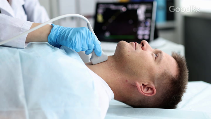
Ultrasound is the imaging modality of choice when evaluating the thyroid. It is a safe, painless, and non-invasive procedure. The procedure uses high-frequency sound waves to produce images of the thyroid and other organs inside your body. Ultrasound provides excellent anatomic detail and plays a vital role in current clinical practice; with accurate diagnosis ultrasound significantly improves the management of numerous medical conditions. The thyroid is a gland situated at the front of the neck. It consists of two lobes that lay on either side of the windpipe (trachea) and are connected by a strip of thyroid tissue called the isthmus. The thyroid gland is an endocrine gland that produces and releases hormones into the bloodstream.
What happens during my scan?
A thyroid ultrasound scan doesn’t need preparation. You just have to expose your neck. You will be examined in a semi-sitting position with your chin upwards. A high-frequency linear transducer array will be used to provide a high-resolution image. Greyscale and color doppler ultrasound are used to evaluate a thyroid lesion. The size, form, borders, echogenicity, contents, location, and vascular pattern of the whole thyroid gland will be assessed. In some cases, fine needle aspiration or biopsy might be recommended when further investigations are needed.
Can ultrasound detect any nodules in my thyroid?
Ultrasound has become an essential diagnostic technique in the examination of thyroid nodules. It is highly sensitive in detecting nodules, the sonographic features, and whether they are benign nodules or if they have malignant characteristics that require further investigation such as a biopsy or FNA. Fine needle aspiration (FNA) or Biopsy can help to improve the accuracy of diagnosis.
What is a thyroid nodule?
Thyroid nodules are categorized according to the “U” classification by the British thyroid association. Under this classification, Nodules are classified into categories (U1-U5) based on features including echogenicity, margins, internal echo pattern, calcification, and vascularity. (U1) indicates a normal thyroid. A benign thyroid nodule is indicated by (U2). A U3 nodule is indeterminate/equivocal. (U4) indicates a suspicious nodule and (U5) indicates malignancy. U3-U5 nodules require FNA or Biopsy to be performed.
What else can be detected in my thyroid scan?
Thyroid ultrasound is also used to evaluate diffuse changes in thyroid parenchyma. Hashimoto thyroiditis and Graves’ disease are common disorders that present diffuse thyroid enlargement. Hashimoto thyroiditis is an autoimmune disease where the immune system attacks the thyroid gland therefore the thyroid is unable to produce enough hormones. Graves’ disease is another autoimmune disorder that causes the thyroid to become overactive and produce excess thyroid hormones thus several body functions work faster than usual.

